
Mouse anti-CDKN1B Monoclonal Antibody(1373CT407.103.103)描述别名宿主特异性反应种属应用分子量类型克隆号同种型储存/保存方法研究领域背景说明细胞定位UniProt参考文献
| 概述 | |
| 描述 |
Purified Mouse Monoclonal Antibody (Mab)
|
| 别名 |
CDKN1B抗体;Cyclin-dependent kinase inhibitor 1B; Cyclin-dependent kinase inhibitor p27; p27Kip1; CDKN1B; KIP1
|
| 宿主 |
Mouse
|
| 特异性 |
This CDKN1B antibody is generated from a mouse immunized with CDKN1B recombinant protein.
|
| 反应种属 |
Human
|
| 应用 |
IHC-P~~1:25
WB~~1:1000 |
| 分子量 |
Predicted molecular weight: 22kD
Disclaimer note: The observed molecular weight of the protein may vary from the listed predicted molecular weight due to post translational modifications, post translation cleavages, relative charges, and other experimental factors. |
| 性能 | |
| 类型 |
Monoclonal Antibody
|
| 克隆号 |
1373CT407.103.103
|
| 同种型 |
IgG1,κ
|
| 储存/保存方法 |
Maintain refrigerated at 2-8°C for up to 2 weeks. For long term storage store at -20°C in small aliquots to prevent freeze-thaw cycles.
|
| 研究领域 |
Cancer;Cell Biology
|
| 靶标 | |
| 背景说明 |
Important regulator of cell cycle progression. Involved in G1 arrest. Potent inhibitor of cyclin E- and cyclin A-CDK2 complexes. Forms a complex with cyclin type D-CDK4 complexes and is involved in the assembly, stability, and modulation of CCND1- CDK4 complex activation. Acts either as an inhibitor or an activator of cyclin type D-CDK4 complexes depending on its phosphorylation state and/or stoichometry.
|
| 细胞定位 |
Nucleus. Cytoplasm. Endosome. Note=Nuclear and cytoplasmic in quiescent cells. AKT- or RSK- mediated phosphorylation on Thr-198, binds 14-3-3, translocates to the cytoplasm and promotes cell cycle progression. Mitogen- activated UHMK1 phosphorylation on Ser-10 also results in translocation to the cytoplasm and cell cycle progression Phosphorylation on Ser-10 facilitates nuclear export. Translocates to the nucleus on phosphorylation of Tyr-88 and Tyr-89 Colocalizes at the endosome with SNX6; this leads to lysosomal degradation (By similarity).
|
| UniProt |
P46527
|
| 参考文献 | |
| 参考文献 |
Polyak K.,et al.Cell 78:59-66(1994).
Pietenpol J.A.,et al.Cancer Res. 55:1206-1210(1995). Kalnine N.,et al.Submitted (OCT-2004) to the EMBL/GenBank/DDBJ databases. Montagnoli A.,et al.Genes Dev. 13:1181-1189(1999). Ishida N.,et al.J. Biol. Chem. 275:25146-25154(2000). |
实验结果图

Immunohistochemical analysis of paraffin-embedded H. skeletal muscle section using CDKN1B Antibody(Cat#JP100463). JP100463 was diluted at 1:25 dilution. A peroxidase-conjugated goat anti-mouse IgG at 1:400 dilution was used as the secondary antibody, followed by DAB staining.
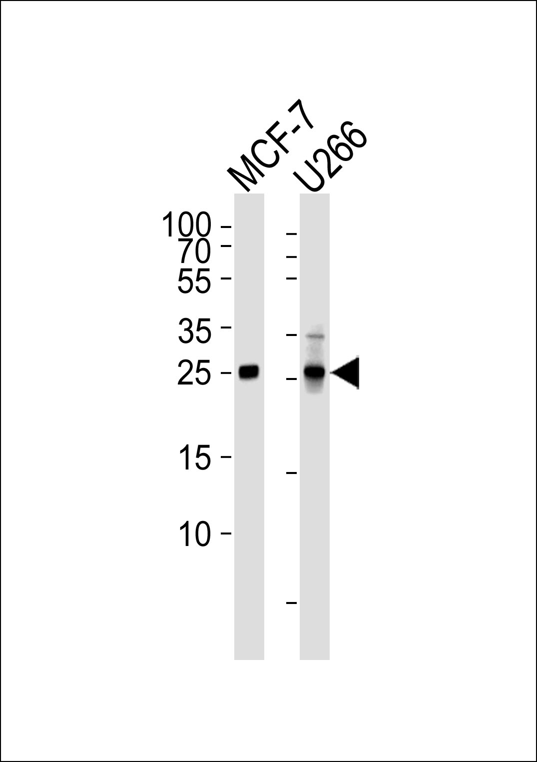
Western blot analysis of lysates from MCF-7, U266 cell line (from left to right), using CDKN1B Antibody(Cat. #JP100463). JP100463 was diluted at 1:1000 at each lane. A goat anti-mouse IgG H&L(HRP) at 1:3000 dilution was used as the secondary antibody. Lysates at 35μg per lane.