
Mouse anti-CHRM2 Monoclonal Antibody(1424CT461.78.60)描述别名宿主特异性反应种属应用分子量类型克隆号同种型储存/保存方法研究领域背景说明细胞定位UniProt参考文献
| 概述 | |
| 描述 |
Purified Mouse Monoclonal Antibody (Mab)
|
| 别名 |
CHRM2抗体;Muscarinic acetylcholine receptor M2; CHRM2
|
| 宿主 |
Mouse
|
| 特异性 |
This antibody is generated from a mouse immunized with a recombinant protein.
|
| 反应种属 |
Human, Mouse
|
| 应用 |
FC~~1:25
IF~~1:25 IHC-P~~1:25 WB~~1:500 |
| 分子量 |
Predicted molecular weight: 52kD
Disclaimer note: The observed molecular weight of the protein may vary from the listed predicted molecular weight due to post translational modifications, post translation cleavages, relative charges, and other experimental factors. |
| 性能 | |
| 类型 |
Monoclonal Antibody
|
| 克隆号 |
1424CT461.78.60
|
| 同种型 |
IgG1,κ
|
| 储存/保存方法 |
Maintain refrigerated at 2-8°C for up to 2 weeks. For long term storage store at -20°C in small aliquots to prevent freeze-thaw cycles.
|
| 研究领域 |
Neuroscience
|
| 靶标 | |
| 背景说明 |
The muscarinic acetylcholine receptor mediates various cellular responses, including inhibition of adenylate cyclase, breakdown of phosphoinositides and modulation of potassium channels through the action of G proteins. Primary transducing effect is adenylate cyclase inhibition. Signaling promotes phospholipase C activity, leading to the release of inositol trisphosphate (IP3); this then triggers calcium ion release into the cytosol.
|
| 细胞定位 |
Cell membrane; Multi-pass membrane protein. Cell junction, synapse, postsynaptic cell membrane; Multi-pass membrane protein. Note=Phosphorylation in response to agonist binding promotes receptor internalization.
|
| UniProt |
P08172
|
| 参考文献 | |
| 参考文献 |
Bonner T.I.,et al.Science 237:527-532(1987).
Peralta E.G.,et al.EMBO J. 6:3923-3929(1987). Puhl H.L. III,et al.Submitted (APR-2002) to the EMBL/GenBank/DDBJ databases. Kitano T.,et al.Mol. Biol. Evol. 21:936-944(2004). Gurevich V.V.,et al.J. Biol. Chem. 270:720-731(1995). |
实验结果图
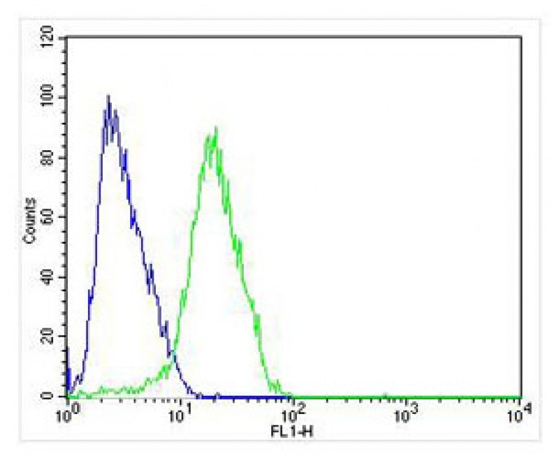
Overlay histogram showing SH-SY5Y cells stained with (green line). The cells were fixed with 4% paraformaldehyde (10 min) and then permeabilized with 90% methanol for 10 min. The cells were then icubated in 2% bovine serum albumin to block non-specific protein-protein interactions followed by the antibody (, 1:25 dilution) for 60 min at 37ºC. The secondary antibody used was Alexa Fluor® 488 goat anti-mouse lgG (166821) at 1/200 dilution for 40 min at 37ºC. Isotype control antibody (blue line) was mouse IgG1 (1μg/1×10^6 cells) used under the same conditions. Acquisition of >10, 000 events was performed.
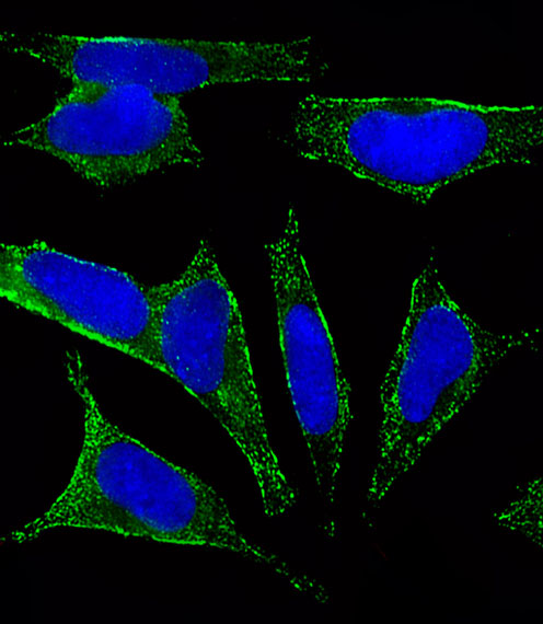
Fluorescent image of SH-SY5Y cells stained with CHRM2 Antibody (Cat#JP100476 ). JP100476 was diluted at 1:25 dilution. An Alexa Fluor® 488-conjugated goat anti-mouse lgG at 1:400 dilution was used as the secondary antibody (green). DAPI was used to stain the cell nuclear (blue).
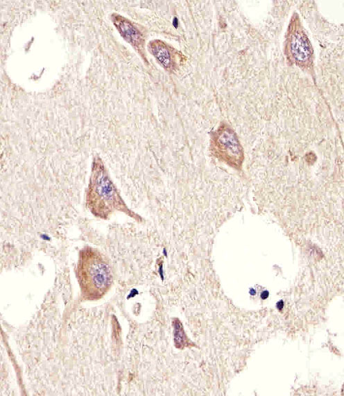
Immunohistochemical analysis of paraffin-embedded H. brain section using CHRM2(Cat#JP100476 ). JP100476 was diluted at 1:25 dilution. A undiluted biotinylated goat polyvalent antibody was used as the secondary, followed by DAB staining.
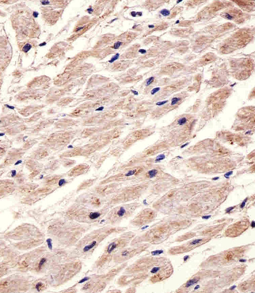
Immunohistochemical analysis of paraffin-embedded H. heart section using CHRM2 (Cat#JP100476 ). JP100476 was diluted at 1:25 dilution. A undiluted biotinylated goat polyvalent antibody was used as the secondary, followed by DAB staining.
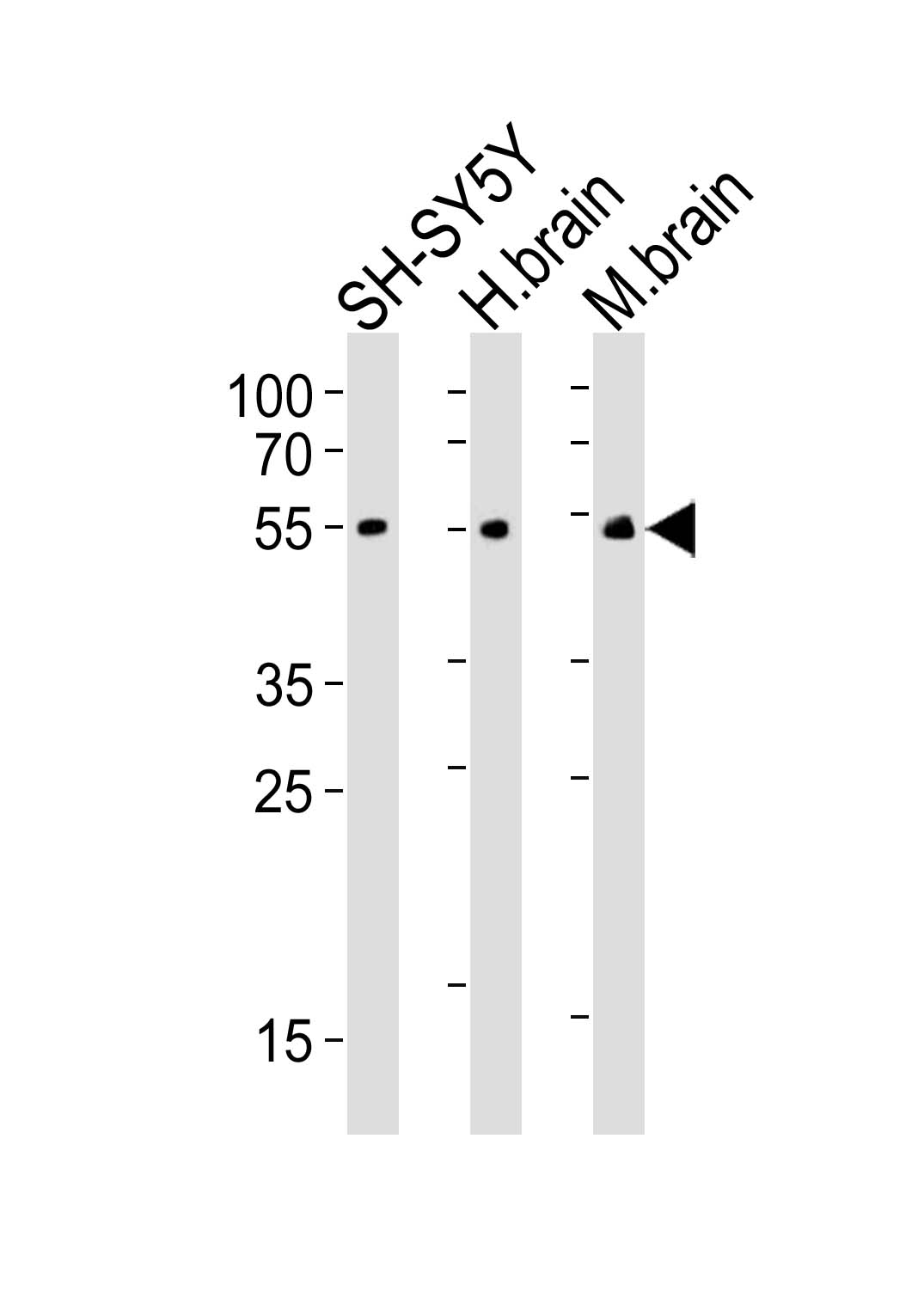
Western blot analysis of lysates from SH-SY5Y cell line, human brain, mouse brain tissue(from left to right), using CHRM2 Antibody(Cat. #JP100476). JP100476 was diluted at 1:500 at each lane. A goat anti-mouse IgG H&L(HRP) at 1:3000 dilution was used as the secondary antibody. Lysates at 20μg per lane.