
Rabbit anti-Calponin-1 Monoclonal Antibody(080-78)别名宿主反应种属应用分子量免疫原形式浓度纯化方法类型克隆号储存/保存方法存储溶液背景说明UniProt
| 概述 | |
| 别名 |
Basic calponin; Calponin H1 (smooth muscle); CNN1
|
| 宿主 |
Rabbit
|
| 反应种属 |
Human, Mouse, Rat
|
| 应用 |
WB: 1:1000,IHC: 1:2000,ICC: 1: 250
|
| 分子量 |
34kDa
|
| 免疫原 |
Synthetic peptide
|
| 性能 | |
| 形式 |
liquid
|
| 浓度 |
0.5 mg/ml
|
| 纯化方法 |
Protein A affinity column
|
| 类型 |
Monoclonal antibody
|
| 克隆号 |
080-78
|
| 储存/保存方法 |
Store at -20℃ for one year.
|
| 存储溶液 |
PBS, 40% Glycerol, 0.05%BSA, 0.03% Proclin 300
|
| 靶标 | |
| 背景说明 |
Calponin-1 is a 34 kDa actin-binding protein with regions of sequence homology to cardiac troponin I and T. It is located in the cytoskeleton and contractile apparatus of differentiated smooth muscle cells. There are 3 isoforms of calponin: calponin-1 (h1 or basic), calponin-2 (h2 or neutral), and calponin-3 (h3 or acidic). Calponin-1 is implicated in the regulation of smooth muscle contraction by mediating intracellular signaling responses to some vasoconstrictors by acting as a contractile scaffold protein. This mechanism is based on results in aortic smooth muscle cells stimulated with phenylephrine, which showed calponin-1 connecting protein kinase C and ERK1/2 pathways to promote contractility.
|
| UniProt |
P51911
|
实验结果图
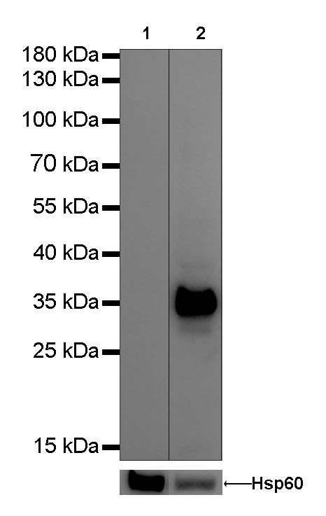
WB result of Calponin-1 Rabbit mAb Primary antibody : Calponin-1 Rabbit mAb at 1/1000 dilution Lane 1: 293-T whole cell lysate 20 µg Lane 2: mouse bladder lysate 20 µg Negative control: 293-T whole cell lysate Secondary antibody: #JP20040 at 1/10000 dilution Predicted MW: 34 kDa Observed MW: 34 kDa Exposure time: 0.3s

IHC shows positive staining in paraffin-embedded human prostate. Anti-Calponin-1 antibody was used at 1/2000 dilution, Secondary antibody: #JP20040 Counterstained with hematoxylin. Heat mediated antigen retrieval with Tris/EDTA buffer pH9.0 was performed before commencing with IHC staining protocol.

IHC shows positive staining in paraffin-embedded human colon. Anti-Calponin-1 antibody was used at 1/2000 dilution, Secondary antibody: #JP20040 Counterstained with hematoxylin. Heat mediated antigen retrieval with Tris/EDTA buffer pH9.0 was performed before commencing with IHC staining protocol.

IHC shows positive staining in paraffin-embedded human cervix cancer. Anti-Calponin-1 antibody was used at 1/2000 dilution, Secondary antibody: #JP20040 Counterstained with hematoxylin. Heat mediated antigen retrieval with Tris/EDTA buffer pH9.0 was performed before commencing with IHC staining protocol.

IHC shows positive staining in paraffin-embedded mouse stomach. Anti-Calponin-1 antibody was used at 1/2000 dilution, Secondary antibody: #JP20040 Counterstained with hematoxylin. Heat mediated antigen retrieval with Tris/EDTA buffer pH9.0 was performed before commencing with IHC staining protocol.

IHC shows positive staining in paraffin-embedded rat colon. Anti-Calponin-1 antibody was used at 1/2000 dilution, Secondary antibody: #JP20040 Counterstained with hematoxylin. Heat mediated antigen retrieval with Tris/EDTA buffer pH9.0 was performed before commencing with IHC staining protocol.

ICC shows cytoplasm staining in Panc-1 cells. Anti-Calponin-1 antibody was used at 1/250 dilution and incubated overnight at 4°C. Secondary antibody: #JP20025 at 1/1000 dilution. Cells were fixed with 100% Methanol and permeabilized with 0.1% Triton X-100. Nuclei were countersained with DAPI.

Negative control: ICC shows negative staining in 293T cells. Anti-Calponin-1 antibody was used at 1/250 dilution and incubated overnight at 4°C. Secondary antibody: #JP20025 at 1/1000 dilution. Cells were fixed with 100% Methanol and permeabilized with 0.1% Triton X-100. Nuclei were countersained with DAPI.