
Rabbit anti-p53 Recombinant Monoclonal Antibody(100-72)描述别名宿主特异性反应种属应用分子量免疫原形式浓度纯化方法类型克隆号储存/保存方法存储溶液背景说明组织特异性翻译后修饰细胞定位UniProt
| 概述 | |
| 描述 |
Tumor protein p53, a nuclear protein, plays an essential role in the regulation of cell cycle, specifically in the transition from G0 to G1. It is found in very low levels in normal cells, however, in a variety of transformed cell lines, it is expressed in high amounts, and believed to contribute to transformation and malignancy. p53 is a DNA-binding protein containing DNA-binding, oligomerization and transcription activation domains. It is postulated to bind as a tetramer to a p53-binding site and activate expression of downstream genes that inhibit growth and/or invasion, and thus function as a tumor suppressor. Mutants of p53 that frequently occur in a number of different human cancers fail to bind the consensus DNA binding site, and hence cause the loss of tumor suppressor activity. Alterations of the TP53 gene occur not only as somatic mutations in human malignancies, but also as germline mutations in some cancer-prone families with Li-Fraumeni syndrome.
|
| 别名 |
p53抗体;Antigen NY-CO-13; Phosphoprotein p53; Tumor suppressor p53; TP53
|
| 宿主 |
Rabbit
|
| 特异性 |
p53 Antibody detects endogenous levels of p53.
|
| 反应种属 |
Human
|
| 应用 |
WB: 1: 2500, IHC-P: 1:1000, FC(Intra) : 1:250, ICC: 1:500
|
| 分子量 |
53kDa
|
| 免疫原 |
Synthetic peptide
|
| 性能 | |
| 形式 |
liquid
|
| 浓度 |
0.25 mg/ml
|
| 纯化方法 |
Protein A affinity column
|
| 类型 |
Monoclonal antibody
|
| 克隆号 |
100-72
|
| 储存/保存方法 |
Store at -20℃ for one year.
|
| 存储溶液 |
PBS, 40% Glycerol, 0.05% BSA, 0.03% Proclin 300
|
| 靶标 | |
| 背景说明 |
p53, also known as Tumor protein P53, cellular tumor antigen p53 (UniProt name), or transformation-related protein 53 (TRP53) is a regulatory protein that is often mutated in human cancers. The p53 proteins (originally thought to be, and often spoken of as, a single protein) are crucial in vertebrates, where they prevent cancer formation. As such, p53 has been described as “the guardian of the genome” because of its role in conserving stability by preventing genome mutation. Hence TP53 is classified as a tumor suppressor gene.
|
| 组织特异性 |
Ubiquitous. Isoforms are expressed in a wide range of normal tissues but in a tissue-dependent manner. Isoform 2 is expressed in most normal tissues but is not detected in brain, lung, prostate, muscle, fetal brain, spinal cord and fetal liver. Isoform 3 is expressed in most normal tissues but is not detected in lung, spleen, testis, fetal brain, spinal cord and fetal liver. Isoform 7 is expressed in most normal tissues but is not detected in prostate, uterus, skeletal muscle and breast. Isoform 8 is detected only in colon, bone marrow, testis, fetal brain and intestine. Isoform 9 is expressed in most normal tissues but is not detected in brain, heart, lung, fetal liver, salivary gland, breast or intestine.
|
| 翻译后修饰 |
Acetylated. Acetylation of Lys-382 by CREBBP enhances transcriptional activity. Deacetylation of Lys-382 by SIRT1 impairs its ability to induce proapoptotic program and modulate cell senescence. Deacetylation by SIRT2 impairs its ability to induce transcription activation in a AKT-dependent manner.Phosphorylation on Ser residues mediates transcriptional activation. Phosphorylated by HIPK1 (By similarity). Phosphorylation at Ser-9 by HIPK4 increases repression activity on BIRC5 promoter. Phosphorylated on Thr-18 by VRK1. Phosphorylated on Ser-20 by CHEK2 in response to DNA damage, which prevents ubiquitination by MDM2. Phosphorylated on Ser-20 by PLK3 in response to reactive oxygen species (ROS), promoting p53/TP53-mediated apoptosis. Phosphorylated on Thr-55 by TAF1, which promotes MDM2-mediated degradation. Phosphorylated on Ser-33 by CDK7 in a CAK complex in response to DNA damage. Phosphorylated on Ser-46 by HIPK2 upon UV irradiation. Phosphorylation on Ser-46 is required for acetylation by CREBBP. Phosphorylated on Ser-392 following UV but not gamma irradiation. Phosphorylated on Ser-15 upon ultraviolet irradiation; which is enhanced by interaction with BANP. Phosphorylated by NUAK1 at Ser-15 and Ser-392; was initially thought to be mediated by STK11/LKB1 but it was later shown that it is indirect and that STK11/LKB1-dependent phosphorylation is probably mediated by downstream NUAK1 (PubMed:21317932). It is unclear whether AMP directly mediates phosphorylation at Ser-15. Phosphorylated on Thr-18 by isoform 1 and isoform 2 of VRK2. Phosphorylation on Thr-18 by isoform 2 of VRK2 results in a reduction in ubiquitination by MDM2 and an increase in acetylation by EP300. Stabilized by CDK5-mediated phosphorylation in response to genotoxic and oxidative stresses at Ser-15, Ser-33 and Ser-46, leading to accumulation of p53/TP53, particularly in the nucleus, thus inducing the transactivation of p53/TP53 target genes. Phosphorylated by DYRK2 at Ser-46 in response to genotoxic stress. Phosphorylated at Ser-315 and Ser-392 by CDK2 in response to DNA-damage.Dephosphorylated by PP2A-PPP2R5C holoenzyme at Thr-55. SV40 small T antigen inhibits the dephosphorylation by the AC form of PP2A.May be O-glycosylated in the C-terminal basic region. Studied in EB-1 cell line.Ubiquitinated by MDM2 and SYVN1, which leads to proteasomal degradation (PubMed:10722742, PubMed:12810724, PubMed:15340061, PubMed:17170702, PubMed:19880522). Ubiquitinated by RFWD3, which works in cooperation with MDM2 and may catalyze the formation of short polyubiquitin chains on p53/TP53 that are not targeted to the proteasome (PubMed:10722742, PubMed:12810724, PubMed:20173098). Ubiquitinated by MKRN1 at Lys-291 and Lys-292, which leads to proteasomal degradation (PubMed:19536131). Deubiquitinated by USP10, leading to its stabilization (PubMed:20096447). Ubiquitinated by TRIM24, RFFL, RNF34 and RNF125, which leads to proteasomal degradation (PubMed:19556538). Ubiquitination by TOPORS induces degradation (PubMed:19473992). Deubiquitination by USP7, leading to stabilization (PubMed:15053880). Isoform 4 is monoubiquitinated in an MDM2-independent manner (PubMed:15340061). Ubiquitinated by RFWD2, which leads to proteasomal degradation (PubMed:19837670). Ubiquitination and subsequent proteasomal degradation is negatively regulated by CCAR2 (PubMed:25732823).Monomethylated at Lys-372 by SETD7, leading to stabilization and increased transcriptional activation. Monomethylated at Lys-370 by SMYD2, leading to decreased DNA-binding activity and subsequent transcriptional regulation activity. Lys-372 monomethylation prevents interaction with SMYD2 and subsequent monomethylation at Lys-370. Dimethylated at Lys-373 by EHMT1 and EHMT2. Monomethylated at Lys-382 by KMT5A, promoting interaction with L3MBTL1 and leading to repress transcriptional activity. Dimethylation at Lys-370 and Lys-382 diminishes p53 ubiquitination, through stabilizing association with the methyl reader PHF20. Demethylation of dimethylated Lys-370 by KDM1A prevents interaction with TP53BP1 and represses TP53-mediated transcriptional activation.Sumoylated with SUMO1. Sumoylated at Lys-386 by UBC9.
|
| 细胞定位 |
Nucleus, cytoplasm
|
| UniProt |
P04637
|
实验结果图
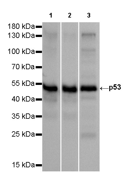
WB result of p53 Rabbit mAb Primary antibody: p53 Rabbit mAb at 1/2500 dilution Lane 1: A431 whole cell lysate 20 µg Lane 2: HT-29 whole cell lysate 20 µg Lane 3: T47D whole cell lysate 20 µg Secondary antibody: #JP20040 at 1/10000 dilution Predicted MW: 53 kDa Observed MW: 53 kDa Exposure time: 10s

IHC shows positive staining in paraffin-embedded human colon cancer. Anti-p53 antibody was used at 1/1000 dilution, Secondary antibody: #JP20040. Counterstained with hematoxylin. Heat mediated antigen retrieval with Tris/EDTA buffer pH9.0 was performed before commencing with IHC staining protocol.

IHC shows positive staining in paraffin-embedded human colon cancer. Anti-p53 antibody was used at 1/1000 dilution, Secondary antibody: #JP20040. Counterstained with hematoxylin. Heat mediated antigen retrieval with Tris/EDTA buffer pH9.0 was performed before commencing with IHC staining protocol.

IHC shows positive staining in paraffin-embedded human lung cancer. Anti-p53 antibody was used at 1/1000 dilution, Secondary antibody: #JP20040. Counterstained with hematoxylin. Heat mediated antigen retrieval with Tris/EDTA buffer pH9.0 was performed before commencing with IHC staining protocol.
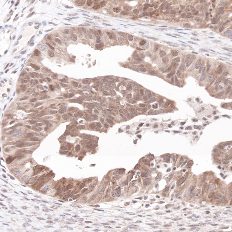
IHC shows positive staining in paraffin-embedded human ovarian cancer. Anti-p53 antibody was used at 1/1000 dilution, Secondary antibody: #JP20040. Counterstained with hematoxylin. Heat mediated antigen retrieval with Tris/EDTA buffer pH9.0 was performed before commencing with IHC staining protocol.
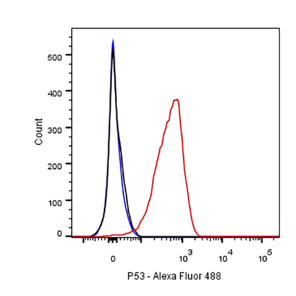
Flow cytometric analysis of HT-29 cells labelling p53 antibody at 1/250 dilution (0.1ug)/ (red) compared with a Rabbit monoclonal IgG (Black) isotype control and an unlabelled control (cells without incubation with primary antibody and secondary antibody) (Blue). Secondary antibody: #JP20025 was used as the secondary antibody.

ICC shows positive nuclear staining in HT-29 cells. Anti-p53 antibody was used at 1/500 dilution and incubated overnight at 4°C. Secondary antibody: #JP20025 at 1/1000 dilution.The cells were fixed with 100% methanol and permeabilized with 0.1% PBS-Triton X-100. Nuclei were countersained with DAPI.