
Rabbit anti-Annexin A2 Recombinant Monoclonal Antibody(444-67)别名宿主反应种属应用免疫原形式浓度纯化方法类型克隆号储存/保存方法存储溶液背景说明细胞定位UniProt
| 概述 | |
| 别名 |
Annexin II; Annexin-2; ANXA2; Calpactin I heavy chain; Calpactin-1 heavy chain; Chromobindin-8; Lipocortin II; Placental anticoagulant protein IV; PAP-IV; Protein I; p36
|
| 宿主 |
Rabbit
|
| 反应种属 |
Human, Mouse, Rat
|
| 应用 |
WB: 1:5000, IHC-P: 1:500
|
| 免疫原 |
Recombinant protein
|
| 性能 | |
| 形式 |
Liquid
|
| 浓度 |
0.5 mg/mL
|
| 纯化方法 |
Protein A affinity column
|
| 类型 |
Monoclonal Antibody
|
| 克隆号 |
444-67
|
| 储存/保存方法 |
Store at -20℃ for one year.
|
| 存储溶液 |
PBS, 40% Glycerol, 0.05% BSA, 0.03% Proclin 300
|
| 靶标 | |
| 背景说明 |
Annexin A2 is a membrane scaffolding and binding protein, which mediated various cellular events. Its functions are generally affected by cellular localization. In the cytoplasm, they interacted with different phospholipid membranes in Ca2+ -dependent manner and play vital roles including actin binding, remodeling and dynamics, cytoskeletal rearrangement, and lipid-raft microdomain formation. However, upon cell exposure to certain stimuli, annexin A2 translocates to the external leaflets of the plasma membrane where annexin A2 was recently reported to serve as a virus receptor, play an important role in the formation of virus replication complex, or implicated in virus assembly and budding [PMID: 30746844].
|
| 细胞定位 |
Secreted, Melanosome, Extracellular matrix
|
| UniProt |
P07355
|
实验结果图

WB result of Annexin A2 Rabbit mAb Primary antibody: Annexin A2 Rabbit mAb at 1/5000 dilution Lane 1: LNCaP whole cell lysate 20 ug Lane 2: HeLa whole cell lysate 20 ug Lane 3: HepG2 whole cell lysate 20 ug Lane 4: K-562 whole cell lysate 20 ug Negative control: LNCaP whole cell lysate Secondary antibody: Goat Anti-Rabbit IgG, (H+L), HRP conjugated at 1/10000 dilution Predicted MW: 39 kDa Observed MW: 35 kDa Exposure time: 10 s

WB result of Annexin A2 Rabbit mAb Primary antibody: Annexin A2 Rabbit mAb at 1/5000 dilution Lane 1: mouse heart lysate 20 ug Secondary antibody: Goat Anti-Rabbit IgG, (H+L), HRP conjugated at 1/10000 dilution Predicted MW: 39 kDa Observed MW: 35 kDa Exposure time: 90 s

WB result of Annexin A2 Rabbit mAb Primary antibody: Annexin A2 Rabbit mAb at 1/5000 dilution Lane 1: rat heart lysate 20 ug Secondary antibody: Goat Anti-Rabbit IgG, (H+L), HRP conjugated at 1/10000 dilution Predicted MW: 39 kDa Observed MW: 35 kDa Exposure time: 90 s

IHC shows positive staining in paraffin-embedded human liver. Anti-Annexin A2 antibody was used at 1/500 dilution, followed by a HRP Polymer for Mouse & Rabbit IgG (ready to use). Counterstained with hematoxylin. Heat mediated antigen retrieval with Tris/EDTA buffer pH9.0 was performed before commencing with IHC staining protocol.

IHC shows positive staining in paraffin-embedded human placenta. Anti-Annexin A2 antibody was used at 1/500 dilution, followed by a HRP Polymer for Mouse & Rabbit IgG (ready to use). Counterstained with hematoxylin. Heat mediated antigen retrieval with Tris/EDTA buffer pH9.0 was performed before commencing with IHC staining protocol.

IHC shows positive staining in paraffin-embedded human stomach. Anti-Annexin A2 antibody was used at 1/500 dilution, followed by a HRP Polymer for Mouse & Rabbit IgG (ready to use). Counterstained with hematoxylin. Heat mediated antigen retrieval with Tris/EDTA buffer pH9.0 was performed before commencing with IHC staining protocol.

IHC shows positive staining in paraffin-embedded human prostate. Anti-Annexin A2 antibody was used at 1/500 dilution, followed by a HRP Polymer for Mouse & Rabbit IgG (ready to use). Counterstained with hematoxylin. Heat mediated antigen retrieval with Tris/EDTA buffer pH9.0 was performed before commencing with IHC staining protocol.

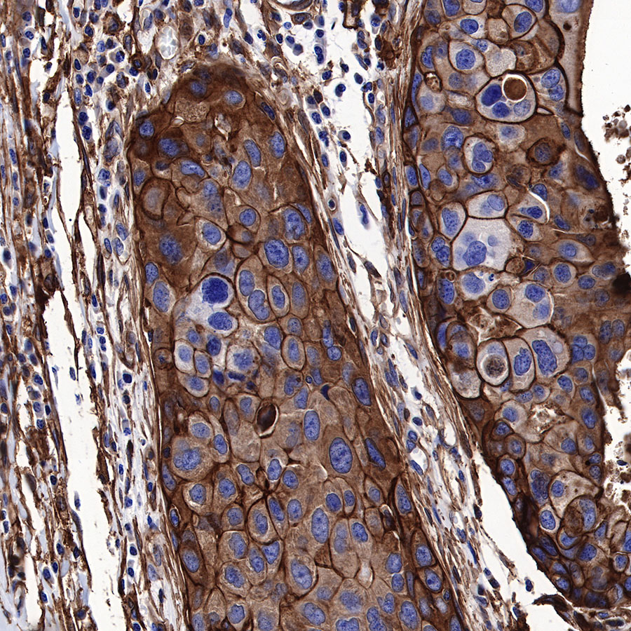
IHC shows positive staining in paraffin-embedded human lung squamous cell carcinoma. Anti-Annexin A2 antibody was used at 1/500 dilution, followed by a HRP Polymer for Mouse & Rabbit IgG (ready to use). Counterstained with hematoxylin. Heat mediated antigen retrieval with Tris/EDTA buffer pH9.0 was performed before commencing with IHC staining protocol.
IHC shows positive staining in paraffin-embedded human breast cancer. Anti-Annexin A2 antibody was used at 1/500 dilution, followed by a HRP Polymer for Mouse & Rabbit IgG (ready to use). Counterstained with hematoxylin. Heat mediated antigen retrieval with Tris/EDTA buffer pH9.0 was performed before commencing with IHC staining protocol.

IHC shows positive staining in paraffin-embedded mouse colon. Anti-Annexin A2 antibody was used at 1/500 dilution, followed by a HRP Polymer for Mouse & Rabbit IgG (ready to use). Counterstained with hematoxylin. Heat mediated antigen retrieval with Tris/EDTA buffer pH9.0 was performed before commencing with IHC staining protocol.

IHC shows positive staining in paraffin-embedded mouse kidney. Anti-Annexin A2 antibody was used at 1/500 dilution, followed by a HRP Polymer for Mouse & Rabbit IgG (ready to use). Counterstained with hematoxylin. Heat mediated antigen retrieval with Tris/EDTA buffer pH9.0 was performed before commencing with IHC staining protocol.

IHC shows positive staining in paraffin-embedded rat colon. Anti-Annexin A2 antibody was used at 1/500 dilution, followed by a HRP Polymer for Mouse & Rabbit IgG (ready to use). Counterstained with hematoxylin. Heat mediated antigen retrieval with Tris/EDTA buffer pH9.0 was performed before commencing with IHC staining protocol.