
Rabbit anti-PEG10 Recombinant Monoclonal Antibody(442-60)别名宿主反应种属应用免疫原形式浓度纯化方法类型克隆号储存/保存方法存储溶液背景说明细胞定位UniProt
| 概述 | |
| 别名 |
Retrotransposon-derived protein PEG10; Embryonal carcinoma differentiation-regulated protein; Mammalian retrotransposon-derived protein 2; MEF3-like protein 1; Paternally expressed gene 10 protein; Ty3/Gypsy-like protein; MEF3L1
|
| 宿主 |
Rabbit
|
| 反应种属 |
Human
|
| 应用 |
WB: 1:1000, IHC-P: 1:1000, ICC: 1:500
|
| 免疫原 |
Recombinant protein
|
| 性能 | |
| 形式 |
Liquid
|
| 浓度 |
0.5 mg/mL
|
| 纯化方法 |
Protein A affinity column
|
| 类型 |
Monoclonal Antibody
|
| 克隆号 |
442-60
|
| 储存/保存方法 |
Store at -20℃ for one year.
|
| 存储溶液 |
PBS, 40% Glycerol, 0.05% BSA, 0.03% Proclin 300
|
| 靶标 | |
| 背景说明 |
Retrotransposon-derived protein PEG10 is a protein that in humans is encoded by the PEG10 gene. Binds its own mRNA in the 5′-UTR region, in the region near the boundary between the nucleocapsid (NC) and protease (PRO) coding sequences and in the beginning of the 3′-UTR region [PubMed:34413232].
|
| 细胞定位 |
Cytoplasm, Nucleus
|
| UniProt |
Q86TG7
|
实验结果图
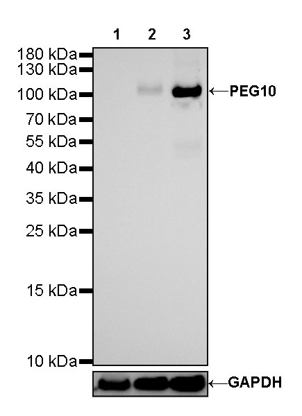
WB result of PEG10 Rabbit mAb Primary antibody: PEG10 Rabbit mAb at 1/1000 dilution Lane 1: 293T whole cell lysate 20 ug Lane 2: HeLa whole cell lysate 20 ug Lane 3: HepG2 whole cell lysate 20 ug Negative control: 293T whole cell lysate Secondary antibody: Goat Anti-Rabbit IgG, (H+L), HRP conjugated at 1/10000 dilution Predicted MW: 80 kDa Observed MW: 110 kDa Exposure time: 180 s
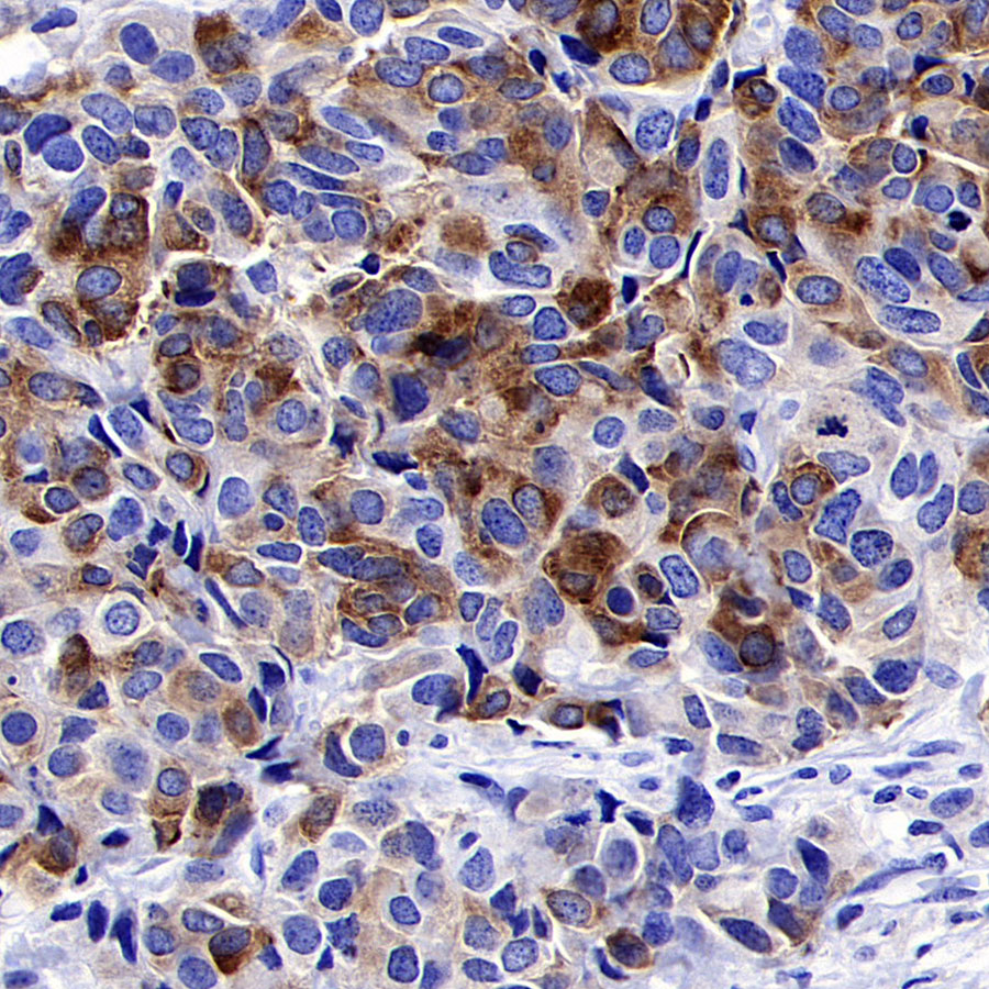
IHC shows positive staining in paraffin-embedded human breast carcinoma. Anti-PEG10 antibody was used at 1/1000 dilution, followed by a HRP Polymer for Mouse & Rabbit IgG (ready to use). Counterstained with hematoxylin. Heat mediated antigen retrieval with Tris/EDTA buffer pH9.0 was performed before commencing with IHC staining protocol.
Negative control: IHC shows negative staining in paraffin-embedded human breast. Anti-PEG10 antibody was used at 1/1000 dilution, followed by a HRP Polymer for Mouse & Rabbit IgG (ready to use). Counterstained with hematoxylin. Heat mediated antigen retrieval with Tris/EDTA buffer pH9.0 was performed before commencing with IHC staining protocol.
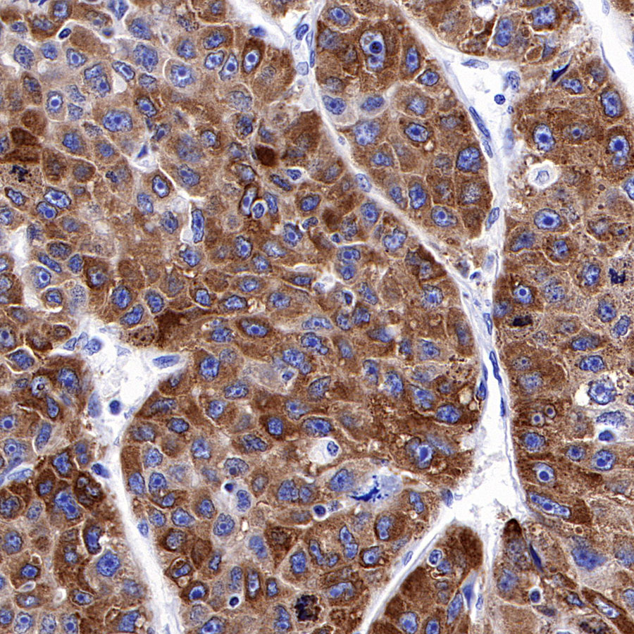
IHC shows positive staining in paraffin-embedded human hepatocellular carcinoma. Anti-PEG10 antibody was used at 1/1000 dilution, followed by a HRP Polymer for Mouse & Rabbit IgG (ready to use). Counterstained with hematoxylin. Heat mediated antigen retrieval with Tris/EDTA buffer pH9.0 was performed before commencing with IHC staining protocol.
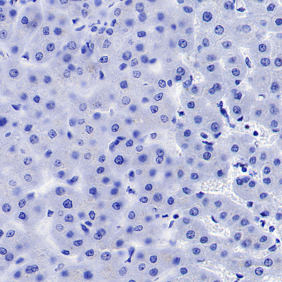
Negative control: IHC shows negative staining in paraffin-embedded human liver. Anti-PEG10 antibody was used at 1/1000 dilution, followed by a HRP Polymer for Mouse & Rabbit IgG (ready to use). Counterstained with hematoxylin. Heat mediated antigen retrieval with Tris/EDTA buffer pH9.0 was performed before commencing with IHC staining protocol.
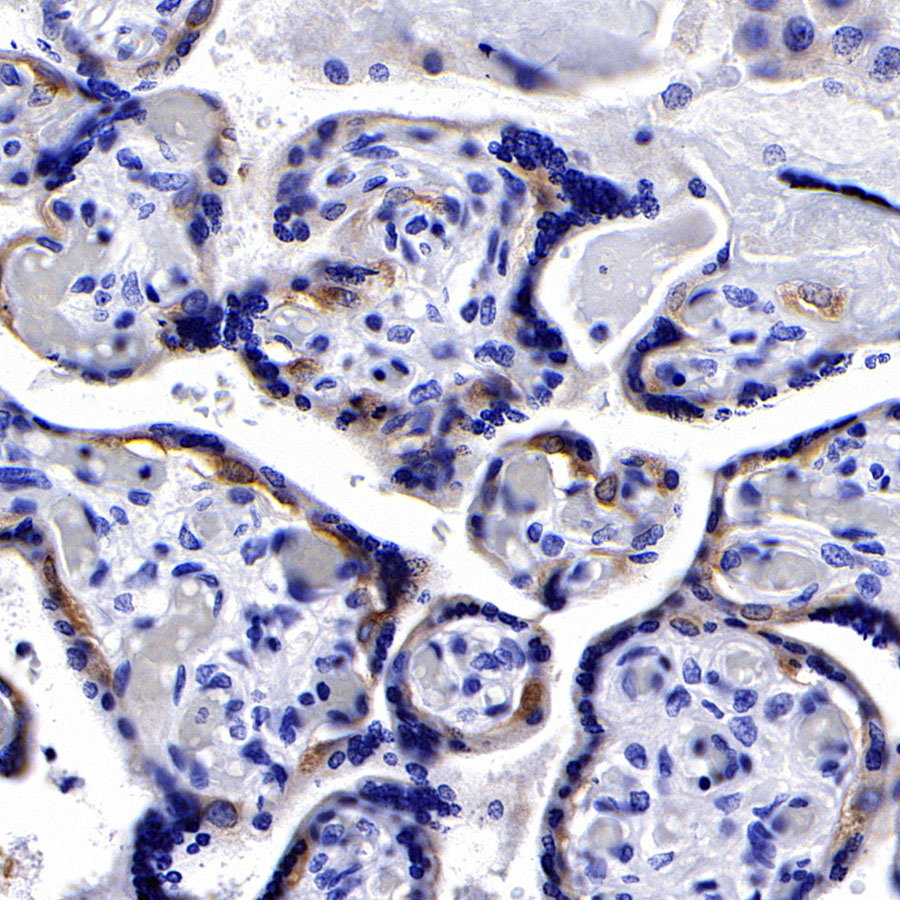
IHC shows positive staining in paraffin-embedded human placenta. Anti-PEG10 antibody was used at 1/1000 dilution, followed by a HRP Polymer for Mouse & Rabbit IgG (ready to use). Counterstained with hematoxylin. Heat mediated antigen retrieval with Tris/EDTA buffer pH9.0 was performed before commencing with IHC staining protocol.
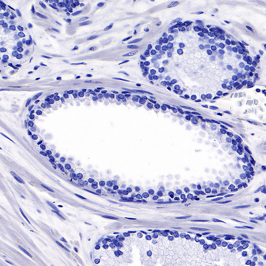
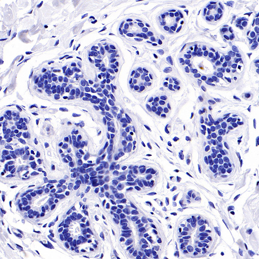
Negative control: IHC shows negative staining in paraffin-embedded human prostate. Anti-PEG10 antibody was used at 1/1000 dilution, followed by a HRP Polymer for Mouse & Rabbit IgG (ready to use). Counterstained with hematoxylin. Heat mediated antigen retrieval with Tris/EDTA buffer pH9.0 was performed before commencing with IHC staining protocol.
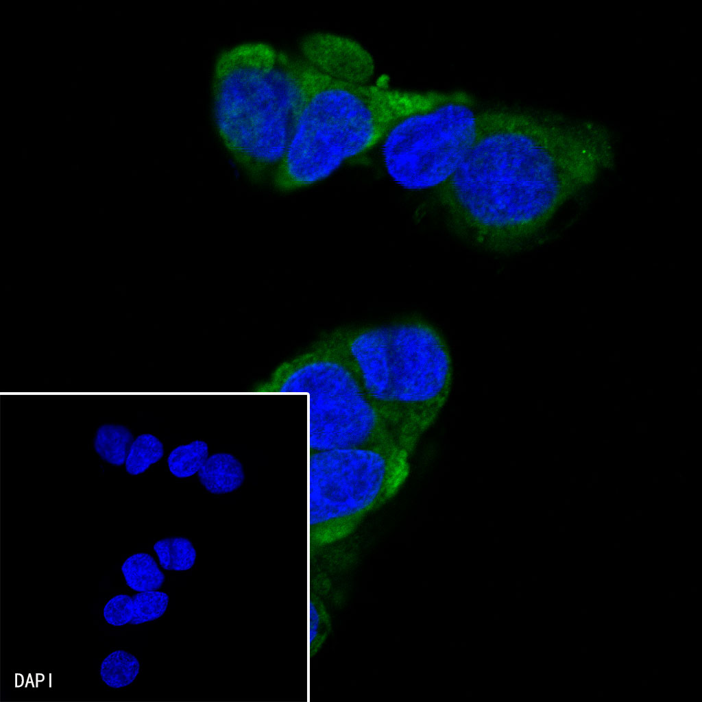
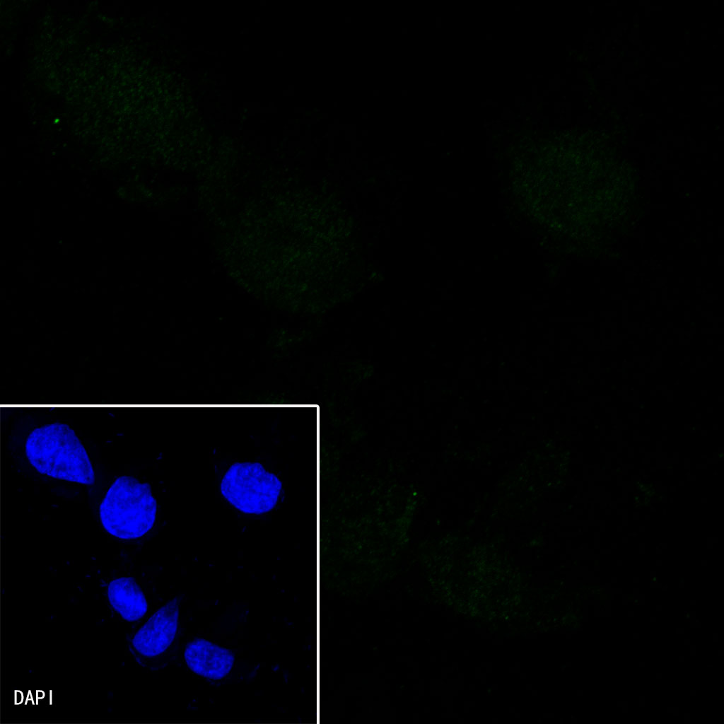
ICC shows positive staining in HepG2 cells. Anti-PEG10 antibody was used at 1/500 dilution (Green) and incubated overnight at 4°C. Goat polyclonal Antibody to Rabbit IgG – H&L (Alexa Fluor® 488) was used as secondary antibody at 1/1000 dilution. The cells were fixed with 4% PFA and permeabilized with 0.1% PBS-Triton X-100. Nuclei were counterstained with DAPI (Blue).
Negative control: ICC shows negative staining in 293T cells. Anti-PEG10 antibody was used at 1/500 dilution and incubated overnight at 4°C. Goat polyclonal Antibody to Rabbit IgG – H&L (Alexa Fluor® 488) was used as secondary antibody at 1/1000 dilution. The cells were fixed with 4% PFA and permeabilized with 0.1% PBS-Triton X-100. Nuclei were counterstained with DAPI (Blue).