
Rabbit anti-LAMP1 Recombinant Monoclonal Antibody(405-39)别名宿主反应种属应用免疫原形式浓度纯化方法类型克隆号储存/保存方法存储溶液背景说明细胞定位UniProt
| 概述 | |
| 别名 |
Lysosome-associated membrane glycoprotein 1; CD107 antigen-like family member A; CD107a
|
| 宿主 |
Rabbit
|
| 反应种属 |
Human
|
| 应用 |
WB: 1:1000, IHC-P: 1:500, ICC: 1:100, FC(Intra): 1:500
|
| 免疫原 |
Recombinant protein
|
| 性能 | |
| 形式 |
Liquid
|
| 浓度 |
0.5 mg/mL
|
| 纯化方法 |
Protein A affinity column
|
| 类型 |
Monoclonal Antibody
|
| 克隆号 |
405-39
|
| 储存/保存方法 |
Store at -20℃ for one year.
|
| 存储溶液 |
PBS, 40% Glycerol, 0.05% BSA, 0.03% Proclin 300
|
| 靶标 | |
| 背景说明 |
Lysosomal-associated membrane protein 1 (LAMP-1) also known as lysosome-associated membrane glycoprotein 1 and CD107a (Cluster of Differentiation 107a), is a protein that in humans is encoded by the LAMP1 gene. The LAMP-1 glycoprotein is a type I transmembrane protein [PMID: 16973206] which is expressed at high or medium levels in at least 76 different normal tissue cell types. It resides primarily across lysosomal membranes [PMID: 2584229], and functions to provide selectins with carbohydrate ligands. LAMP-1 has also been shown to be a marker of degranulation on lymphocytes such as CD8+ and NK cells, and may also play a role in tumor cell differentiation and metastasis.
|
| 细胞定位 |
Cell membrane, Endosome membrane, Lysosome membrane, Late endosome membrane, Cytolytic granule membrane
|
| UniProt |
P11279
|
实验结果图
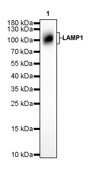
WB result of LAMP1 Rabbit mAb Primary antibody: LAMP1 Rabbit mAb at 1/1000 dilution Lane 1: HeLa whole cell lysate 20 ug Secondary antibody: Goat Anti-Rabbit IgG, (H+L), HRP conjugated at 1/10000 dilution Predicted MW: 45 kDa Observed MW: 90~120 kDa Exposure time: 180 s
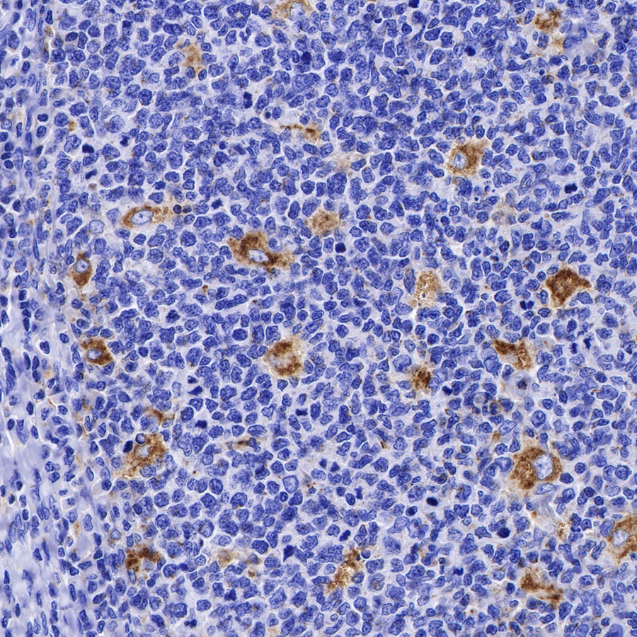
IHC shows positive staining in paraffin-embedded human tonsil. Anti-LAMP1 antibody was used at 1/500 dilution, followed by a HRP Polymer for Mouse & Rabbit IgG (ready to use). Counterstained with hematoxylin. Heat mediated antigen retrieval with Tris/EDTA buffer pH9.0 was performed before commencing with IHC staining protocol.
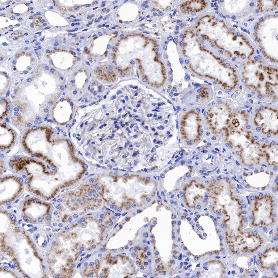
IHC shows positive staining in paraffin-embedded human kidney. Anti-LAMP1 antibody was used at 1/500 dilution, followed by a HRP Polymer for Mouse & Rabbit IgG (ready to use). Counterstained with hematoxylin. Heat mediated antigen retrieval with Tris/EDTA buffer pH9.0 was performed before commencing with IHC staining protocol.
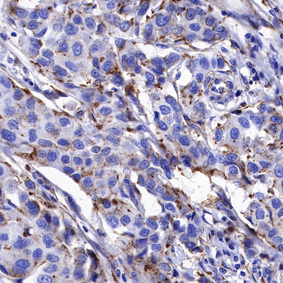
IHC shows positive staining in paraffin-embedded human breast cancer. Anti-LAMP1 antibody was used at 1/500 dilution, followed by a HRP Polymer for Mouse & Rabbit IgG (ready to use). Counterstained with hematoxylin. Heat mediated antigen retrieval with Tris/EDTA buffer pH9.0 was performed before commencing with IHC staining protocol.
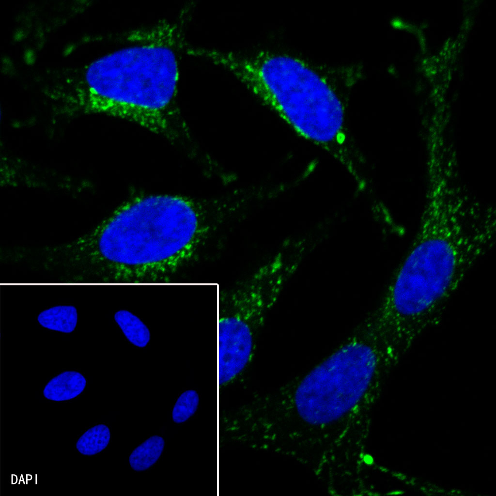
ICC shows positive staining in HeLa cells. Anti-LAMP1 antibody was used at 1/100 dilution (Green) and incubated overnight at 4°C. Goat polyclonal Antibody to Rabbit IgG – H&L (Alexa Fluor® 488) was used as secondary antibody at 1/1000 dilution. The cells were fixed with 100% ice-cold methanol and permeabilized with 0.1% PBS-Triton X-100. Nuclei were counterstained with DAPI (Blue).
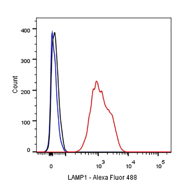
Flow cytometric analysis of HeLa (Human cervix adenocarcinoma epithelial cell) labelling LAMP1 antibody at 1/500 dilution(0.1 μg) / (red) compared with a Rabbit monoclonal IgG (Black) isotype control and an unlabelled control (cells without incubation with primary antibody and secondary antibody) (Blue). Goat Anti-Rabbit IgG Alexa Fluor® 488 was used as the secondary antibody.