
Rabbit anti-Stathmin 1 Recombinant Monoclonal Antibody(383-4)别名宿主反应种属应用免疫原形式浓度纯化方法类型克隆号储存/保存方法存储溶液背景说明细胞定位UniProt
| 概述 | |
| 别名 |
Stathmin; Leukemia-associated phosphoprotein p18; Metablastin; Op18; pp19; Prosolin; Protein Pr22; pp17; C1orf215; LAP18
|
| 宿主 |
Rabbit
|
| 反应种属 |
Human, Mouse, Rat
|
| 应用 |
WB: 1:1000, IHC-P: 1:2000
|
| 免疫原 |
Synthetic Peptide
|
| 性能 | |
| 形式 |
Liquid
|
| 浓度 |
0.5 mg/mL
|
| 纯化方法 |
Protein A affinity column
|
| 类型 |
Monoclonal Antibody
|
| 克隆号 |
383-4
|
| 储存/保存方法 |
Store at -20℃ for one year.
|
| 存储溶液 |
PBS, 40% Glycerol, 0.05% BSA, 0.03% Proclin 300
|
| 靶标 | |
| 背景说明 |
Stathmin 1 (STMN1), also known as p17, p18, p19, 19K, metablastin, oncoprotein 18, LAP 18 and Op18, is a 19 kDa cytosolic protein. It was the first discovered member of a family of phylogenetically related microtubule-destabilizing phosphoproteins critically involved in the construction and function of the mitotic spindle. A threshold level of STMN1 is required for orderly progression through mitosis in a variety of cell types. STMN1 is overexpressed across a broad range of human malignancies (leukemia, lymphoma, neuroblastoma; ovarian, prostatic, breast and lung cancers and mesothelioma). It is also upregulated in normally proliferating cell lines but is only rarely upregulated in nonproliferating cell lines with the exception of neurons, anterior pituitary cells and glial cells [PMID: 18759697].
|
| 细胞定位 |
Cytoplasm, cytoskeleton
|
| UniProt |
P16949
|
实验结果图
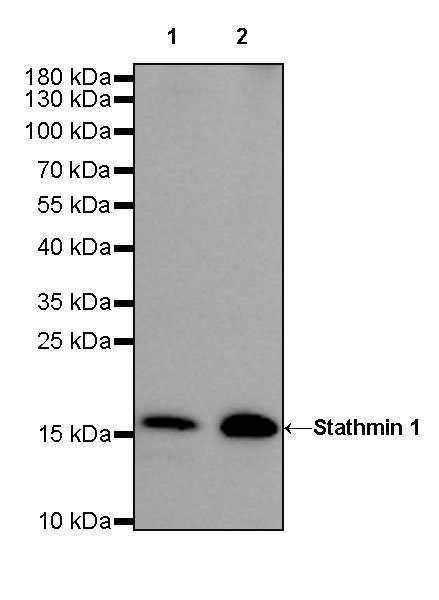
WB result of Stathmin 1 Rabbit mAb Primary antibody: Stathmin 1 Rabbit mAb at 1/1000 dilution Lane 1: MCF7 whole cell lysate 20 µg Lane 2: HeLa whole cell lysate 20 µg Secondary antibody: Goat Anti-Rabbit IgG, (H+L), HRP conjugated at 1/10000 dilution Predicted MW: 17kDa Observed MW: 17kDa Exposure time: 180s
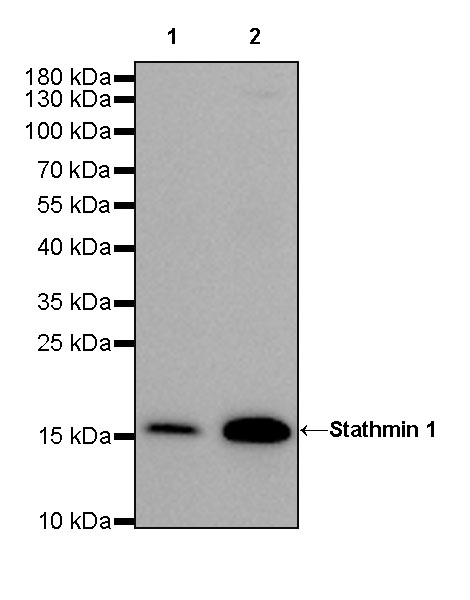
WB result of Stathmin 1 Rabbit mAb Primary antibody: Stathmin 1 Rabbit mAb at 1/1000 dilution Lane 1: NIH/3T3 whole cell lysate 20 µg Lane 2: mouse brain lysate 20 µg Secondary antibody: Goat Anti-Rabbit IgG, (H+L), HRP conjugated at 1/10000 dilution Predicted MW: 17kDa Observed MW: 17kDa Exposure time: 90s
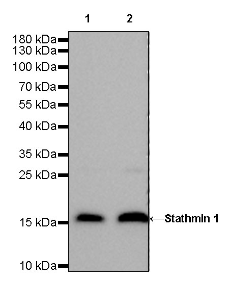
WB result of Stathmin 1 Rabbit mAb Primary antibody: Stathmin 1 Rabbit mAb at 1/1000 dilution Lane 1: C6 whole cell lysate 20 µg Lane 2: rat brain lysate 20 µg Secondary antibody: Goat Anti-Rabbit IgG, (H+L), HRP conjugated at 1/10000 dilution Predicted MW: 17kDa Observed MW: 17kDa Exposure time: 30s
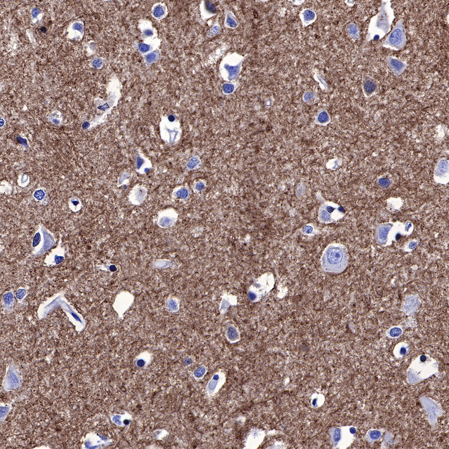
IHC shows positive staining in paraffin-embedded human cerebral cortex. Anti-Stathmin 1 antibody was used at 1/2000 dilution, followed by a HRP Polymer for Mouse & Rabbit IgG (ready to use). Counterstained with hematoxylin. Heat mediated antigen retrieval with Tris/EDTA buffer pH9.0 was performed before commencing with IHC staining protocol.
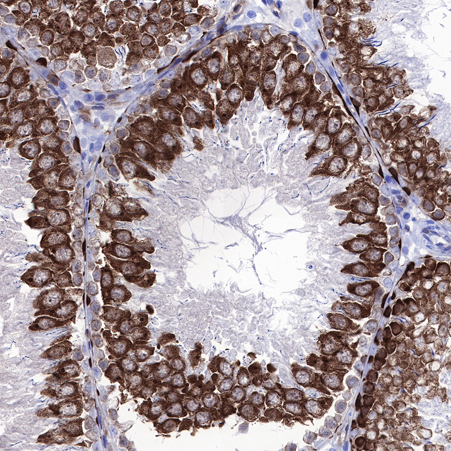
IHC shows positive staining in paraffin-embedded rat testis. Anti-Stathmin 1 antibody was used at 1/2000 dilution, followed by a HRP Polymer for Mouse & Rabbit IgG (ready to use). Counterstained with hematoxylin. Heat mediated antigen retrieval with Tris/EDTA buffer pH9.0 was performed before commencing with IHC staining protocol.
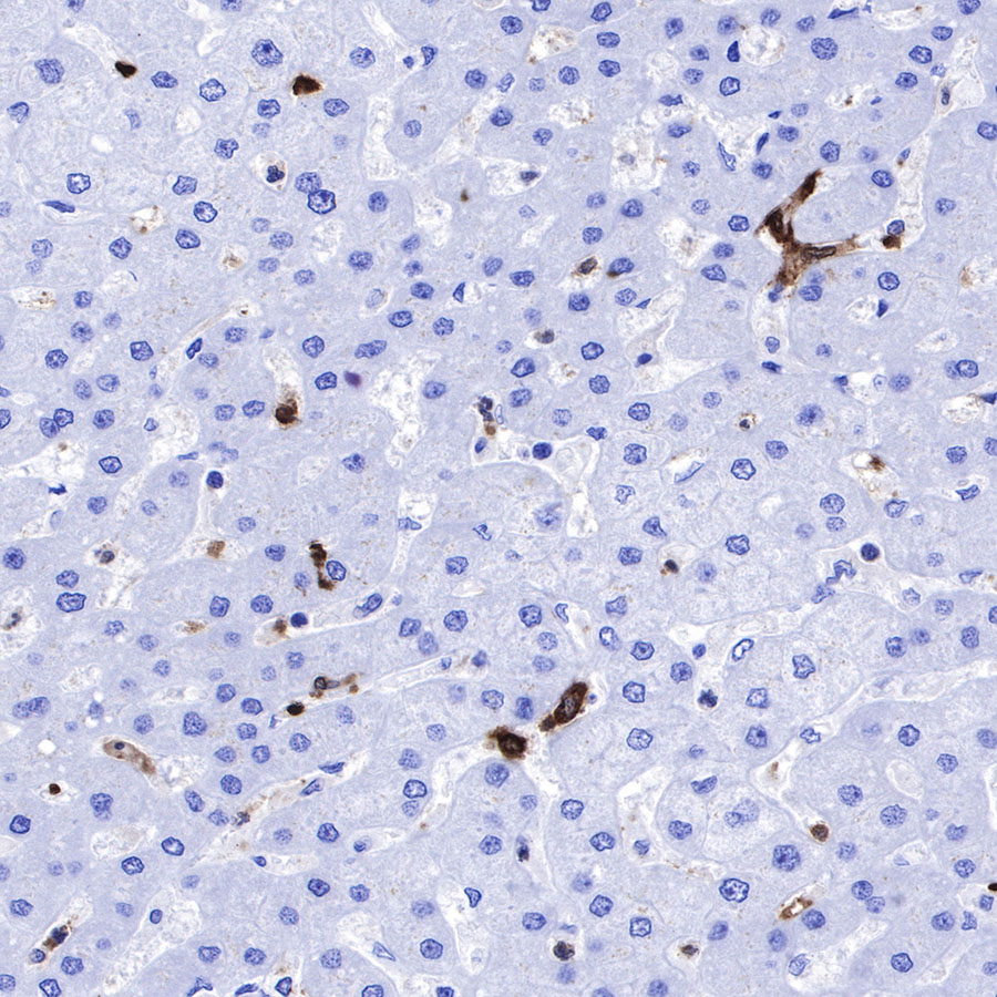
IHC shows positive staining in paraffin-embedded human liver. Anti-Stathmin 1 antibody was used at 1/2000 dilution, followed by a HRP Polymer for Mouse & Rabbit IgG (ready to use). Counterstained with hematoxylin. Heat mediated antigen retrieval with Tris/EDTA buffer pH9.0 was performed before commencing with IHC staining protocol.

IHC shows positive staining in paraffin-embedded human spleen. Anti-Stathmin 1 antibody was used at 1/2000 dilution, followed by a HRP Polymer for Mouse & Rabbit IgG (ready to use). Counterstained with hematoxylin. Heat mediated antigen retrieval with Tris/EDTA buffer pH9.0 was performed before commencing with IHC staining protocol.

IHC shows positive staining in paraffin-embedded human tonsil. Anti-Stathmin 1 antibody was used at 1/2000 dilution, followed by a HRP Polymer for Mouse & Rabbit IgG (ready to use). Counterstained with hematoxylin. Heat mediated antigen retrieval with Tris/EDTA buffer pH9.0 was performed before commencing with IHC staining protocol.
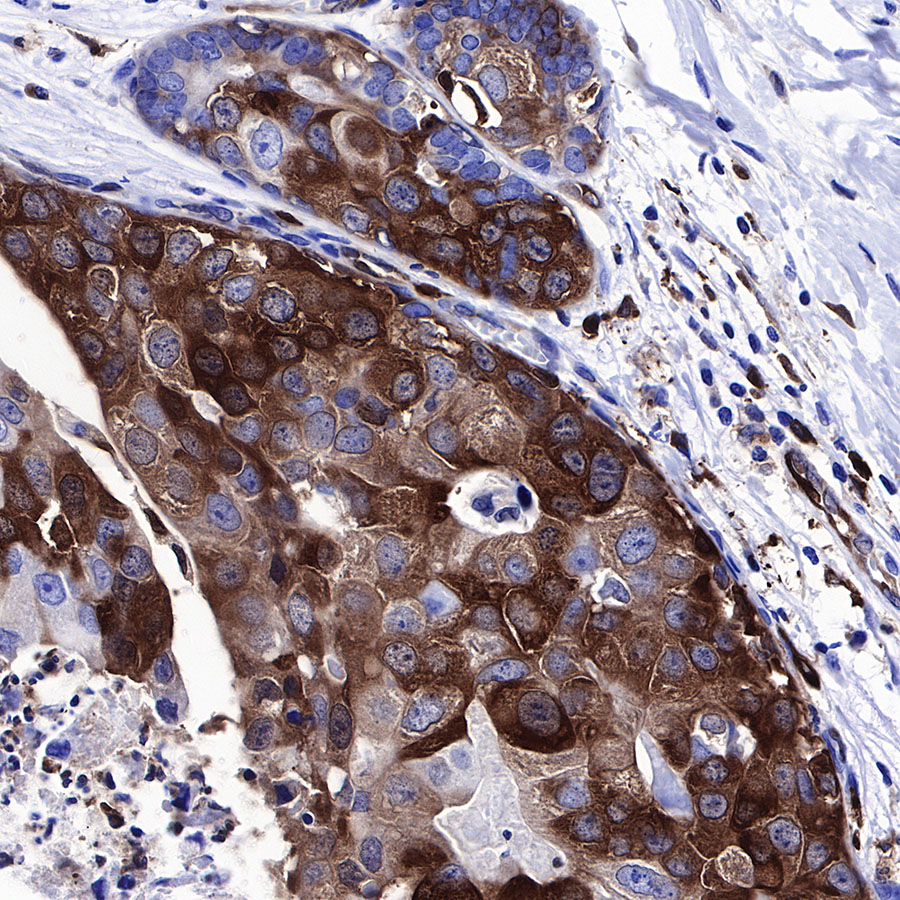
IHC shows positive staining in paraffin-embedded human breast cancer. Anti-Stathmin 1 antibody was used at 1/2000 dilution, followed by a HRP Polymer for Mouse & Rabbit IgG (ready to use). Counterstained with hematoxylin. Heat mediated antigen retrieval with Tris/EDTA buffer pH9.0 was performed before commencing with IHC staining protocol.
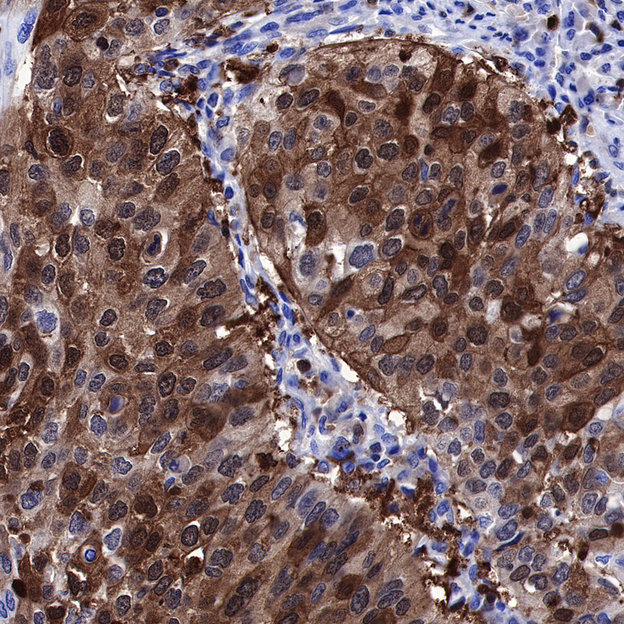
IHC shows positive staining in paraffin-embedded human lung squamous cell carcinoma. Anti-Stathmin 1 antibody was used at 1/2000 dilution, followed by a HRP Polymer for Mouse & Rabbit IgG (ready to use). Counterstained with hematoxylin. Heat mediated antigen retrieval with Tris/EDTA buffer pH9.0 was performed before commencing with IHC staining protocol.

IHC shows positive staining in paraffin-embedded human ovarian carcinoma. Anti-Stathmin 1 antibody was used at 1/2000 dilution, followed by a HRP Polymer for Mouse & Rabbit IgG (ready to use). Counterstained with hematoxylin. Heat mediated antigen retrieval with Tris/EDTA buffer pH9.0 was performed before commencing with IHC staining protocol.

IHC shows positive staining in paraffin-embedded mouse cerebral cortex. Anti-Stathmin 1 antibody was used at 1/2000 dilution, followed by a HRP Polymer for Mouse & Rabbit IgG (ready to use). Counterstained with hematoxylin. Heat mediated antigen retrieval with Tris/EDTA buffer pH9.0 was performed before commencing with IHC staining protocol.
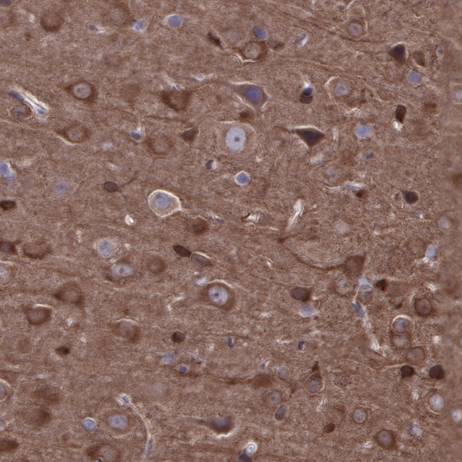
IHC shows positive staining in paraffin-embedded rat cerebral cortex. Anti-Stathmin 1 antibody was used at 1/2000 dilution, followed by a HRP Polymer for Mouse & Rabbit IgG (ready to use). Counterstained with hematoxylin. Heat mediated antigen retrieval with Tris/EDTA buffer pH9.0 was performed before commencing with IHC staining protocol.