
Rabbit anti-CD146 Recombinant Monoclonal Antibody(348-6)别名宿主反应种属应用免疫原形式浓度纯化方法类型克隆号储存/保存方法存储溶液背景说明细胞定位UniProt
| 概述 | |
| 别名 |
MEL-CAM; MUC18; P1H12; A32
|
| 宿主 |
Rabbit
|
| 反应种属 |
Human, Mouse, Rat
|
| 应用 |
WB: 1:1000, IHC-P: 1:1000-1:2000
|
| 免疫原 |
Synthetic peptide
|
| 性能 | |
| 形式 |
Liquid
|
| 浓度 |
0.5 mg/mL
|
| 纯化方法 |
Protein A affinity column
|
| 类型 |
Monoclonal Antibody
|
| 克隆号 |
348-6
|
| 储存/保存方法 |
Store at -20℃ for one year.
|
| 存储溶液 |
PBS, 40% Glycerol, 0.05% BSA, 0.03% Proclin 300
|
| 靶标 | |
| 背景说明 |
Cluster of differentiation 146 (CD146) is a cell adhesion molecule (CAM) which belongs to the immunoglobulin superfamily (IgSF) [PMID: 3542195]. Human CD146 has previously been designated several synonyms, including MUC18 [PMID: 3542195, PMID: 2602381], A32 antigen [PMID: 8162602,PMID: 7943174], S-Endo-1 [PMID: 8573133], melanoma CAM (MCAM or Mel-CAM) [PMID: 7943174, PMID: 8616875, PMID: 9187135], metastasis CAM (MET-CAM) and hemopoietic CAM (HEMCAM). The avian homolog of CD146 has been named gicerin [PMID: 8161457]. CD146 was originally identified as a marker for melanoma (MCAM), due to its overexpression in metastatic lesions and advanced primary tumors, yet not in benign lesions. Increasing amounts of evidence have demonstrated that CD146 is overexpressed in a variety of carcinomas, in addition to melanomas [PMID: 16804906, PMID: 22826148, PMID: 22754372, PMID: 22210108]. As a result of this characteristic, CD146 has attracted attention and is considered to be a potential marker for tumor diagnosis, prognosis and treatment. The majority of studies support the theory that CD146 promotes tumor growth, angiogenesis and metastasis [PMID: 19356677], therefore, CD146 is a promising target for tumor therapy [PMID: 17121934, PMID: 22977083].
|
| 细胞定位 |
Membrane
|
| UniProt |
P43121
|
实验结果图
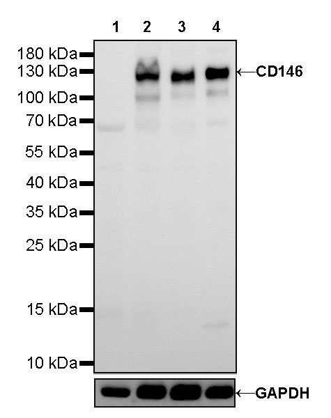
WB result of CD146 Rabbit mAb Primary antibody: CD146 Rabbit mAb at 1/1000 dilution Lane 1: LNCaP whole cell lysate 20 µg Lane 2: SK-MEL-28 whole cell lysate 20 µg Lane 3: HeLa whole cell lysate 20 µg Lane 4: HUVEC whole cell lysate 20 µg Negative control: LNCaP whole cell lysate Secondary antibody: #JP20040 at 1/10000 dilution Predicted MW: 72kDa Observed MW: 120kDa Exposure time: 20s

IHC shows positive staining in paraffin-embedded human melanoma. Anti-CD146 antibody was used at 1/1000 dilution, followed by a HRP Polymer for Mouse & Rabbit IgG (ready to use). Counterstained with hematoxylin. Heat mediated antigen retrieval with Tris/EDTA buffer pH9.0 was performed before commencing with IHC staining protocol.

IHC shows positive staining in paraffin-embedded human melanoma. Anti-CD146 antibody was used at 1/1000 dilution, followed by a HRP Polymer for Mouse & Rabbit IgG (ready to use). Counterstained with hematoxylin. Heat mediated antigen retrieval with Tris/EDTA buffer pH9.0 was performed before commencing with IHC staining protocol.
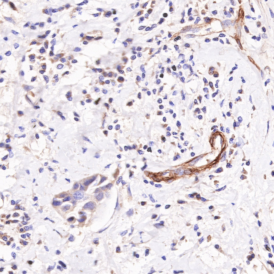
IHC shows positive staining in paraffin-embedded human breast cancer. Anti-CD146 antibody was used at 1/2000 dilution, followed by a HRP Polymer for Mouse & Rabbit IgG (ready to use). Counterstained with hematoxylin. Heat mediated antigen retrieval with Tris/EDTA buffer pH9.0 was performed before commencing with IHC staining protocol.
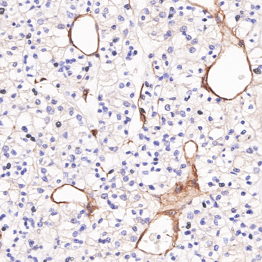
IHC shows positive staining in paraffin-embedded human renal clear cell carcinoma. Anti-CD146 antibody was used at 1/2000 dilution, followed by a HRP Polymer for Mouse & Rabbit IgG (ready to use). Counterstained with hematoxylin. Heat mediated antigen retrieval with Tris/EDTA buffer pH9.0 was performed before commencing with IHC staining protocol.
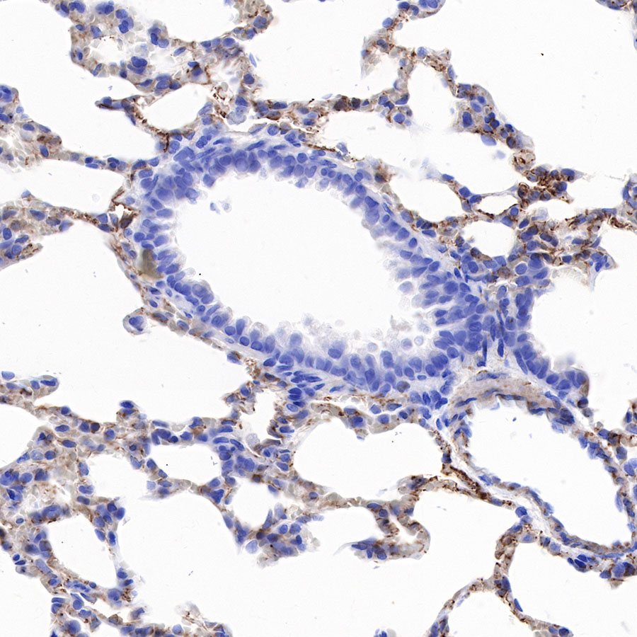
IHC shows positive staining in paraffin-embedded mouse lung. Anti-CD146 antibody was used at 1/2000 dilution, followed by a HRP Polymer for Mouse & Rabbit IgG (ready to use). Counterstained with hematoxylin. Heat mediated antigen retrieval with Tris/EDTA buffer pH9.0 was performed before commencing with IHC staining protocol.
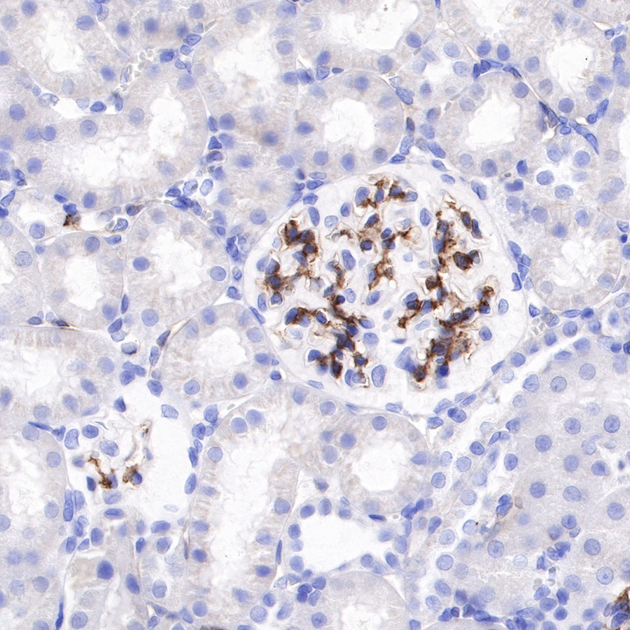
IHC shows positive staining in paraffin-embedded rat kidney. Anti-CD146 antibody was used at 1/2000 dilution, followed by a HRP Polymer for Mouse & Rabbit IgG (ready to use). Counterstained with hematoxylin. Heat mediated antigen retrieval with Tris/EDTA buffer pH9.0 was performed before commencing with IHC staining protocol.