
Rabbit anti-F4/80 Recombinant Monoclonal Antibody(313-49)别名宿主反应种属应用免疫原形式浓度纯化方法类型克隆号储存/保存方法存储溶液背景说明细胞定位UniProt
| 概述 | |
| 别名 |
Adhesion G protein-coupled receptor E1; EGF-like module receptor 1; EMR1 hormone receptor; Gpf480; Adgre1
|
| 宿主 |
Rabbit
|
| 反应种属 |
Mouse, Rat
|
| 应用 |
WB: 1:1000, IHC-P: 1:500
|
| 免疫原 |
Synthetic Peptide
|
| 性能 | |
| 形式 |
Liquid
|
| 浓度 |
0.5 mg/mL
|
| 纯化方法 |
Protein A affinity column
|
| 类型 |
Monoclonal Antibody
|
| 克隆号 |
313-49
|
| 储存/保存方法 |
Store at -20℃ for one year.
|
| 存储溶液 |
PBS, 40% Glycerol, 0.05% BSA, 0.03% Proclin 300
|
| 靶标 | |
| 背景说明 |
The F4/80 monoclonal antibody has been used widely as a marker for mouse macrophages [PMID: 16087400]. EMR1-F4/80 is mainly present in the macrophage-containing stroma-vascular fraction. Furthermore, fibroblasts-like cells (adipoblasts), preadipocytes and adipocytes from the murine cell lines, 3T3-F442A and BFC-1, express CD14 and CD68 mRNA and protein as determined by fluorescence-activated cell sorter, but not F4/80 which, as expected, is strongly expressed in the macrophage cell line RAW264.7 [PMID: 16213494].
|
| 细胞定位 |
Cell membrane
|
| UniProt |
Q61549
|
实验结果图
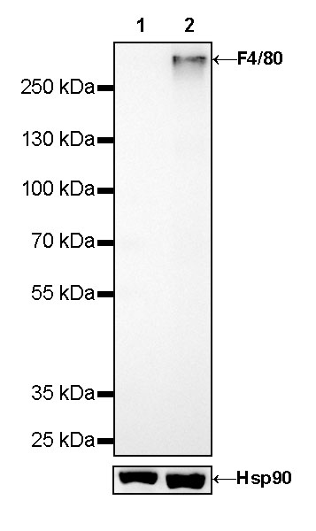
WB result of F4/80 Rabbit mAb Primary antibody: F4/80 Rabbit mAb at 1/1000 dilution Lane 1: NIH/3T3 whole cell lysate 20 µg Lane 2: RAW264.7 whole cell lysate 20 µg Negative control: NIH/3T3 whole cell lysate Secondary antibody: Goat Anti-Rabbit IgG, (H+L), HRP conjugated at 1/10000 dilution Predicted MW: 97kDa Observed MW: 300kDa Exposure time: 180s
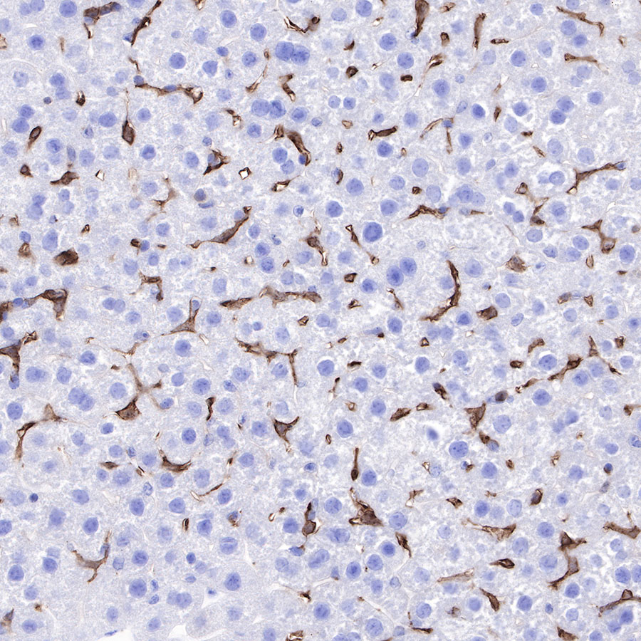
IHC shows positive staining in paraffin-embedded mouse liver. Anti-F4/80 antibody was used at 1/500 dilution, followed by a HRP Polymer for Mouse & Rabbit IgG (ready to use). Counterstained with hematoxylin. Heat mediated antigen retrieval with Tris/EDTA buffer pH9.0 was performed before commencing with IHC staining protocol.
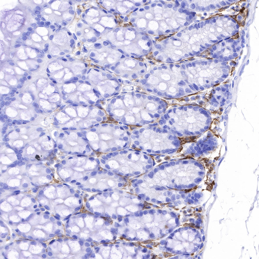
IHC shows positive staining in paraffin-embedded mouse colon. Anti-F4/80 antibody was used at 1/500 dilution, followed by a HRP Polymer for Mouse & Rabbit IgG (ready to use). Counterstained with hematoxylin. Heat mediated antigen retrieval with Tris/EDTA buffer pH9.0 was performed before commencing with IHC staining protocol.
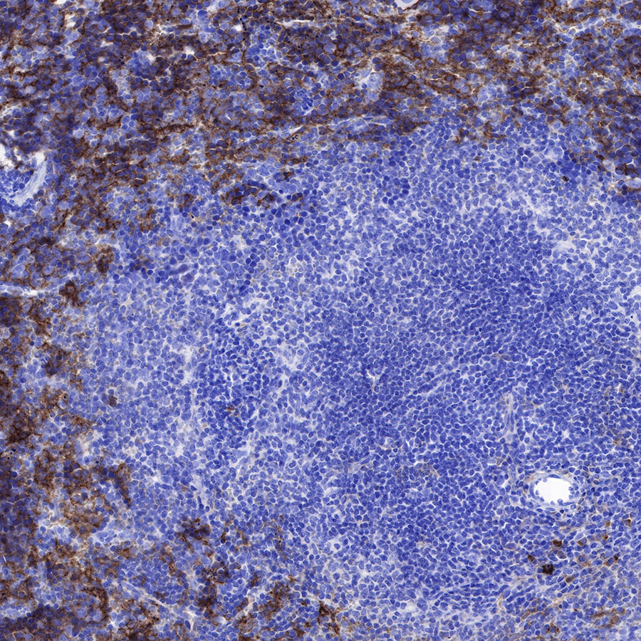
IHC shows positive staining in paraffin-embedded mouse spleen. Anti-F4/80 antibody was used at 1/500 dilution, followed by a HRP Polymer for Mouse & Rabbit IgG (ready to use). Counterstained with hematoxylin. Heat mediated antigen retrieval with Tris/EDTA buffer pH9.0 was performed before commencing with IHC staining protocol.
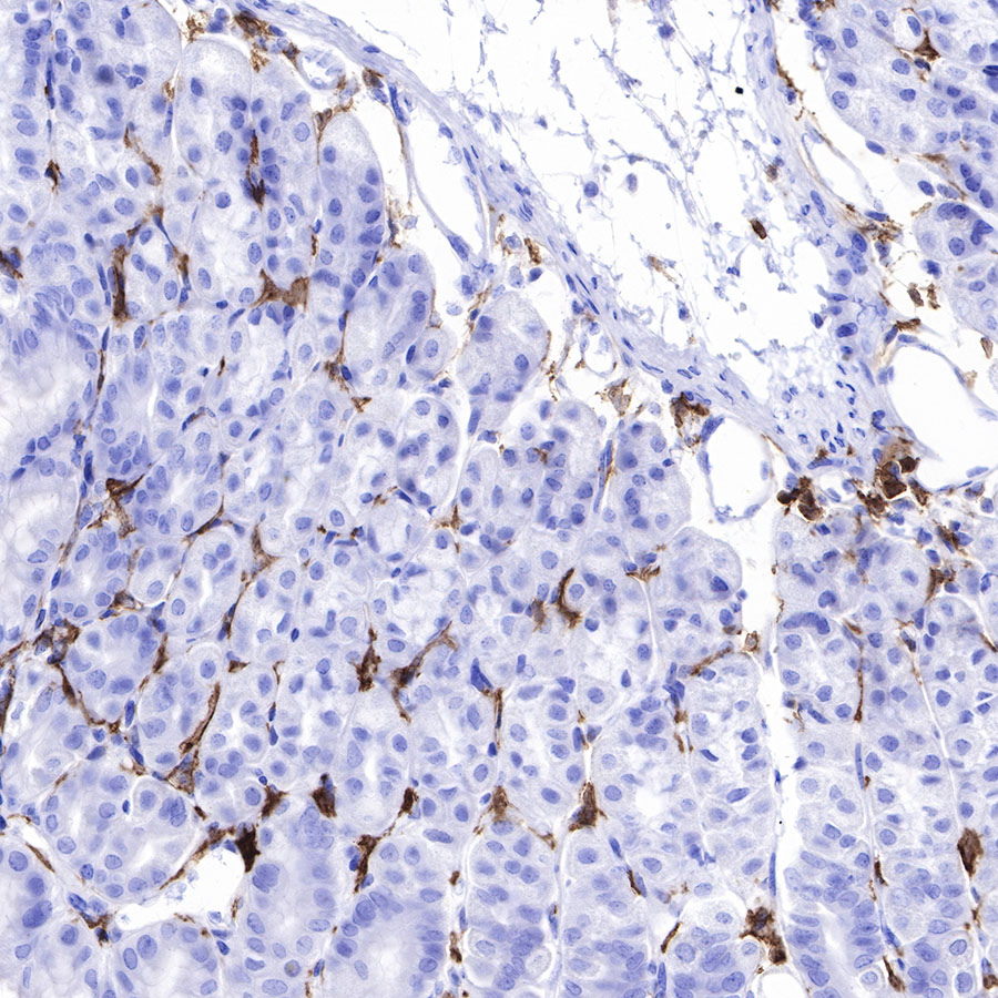
IHC shows positive staining in paraffin-embedded mouse stomach. Anti-F4/80 antibody was used at 1/500 dilution, followed by a HRP Polymer for Mouse & Rabbit IgG (ready to use). Counterstained with hematoxylin. Heat mediated antigen retrieval with Tris/EDTA buffer pH9.0 was performed before commencing with IHC staining protocol.
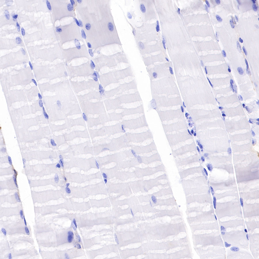
Negative control: IHC shows negative staining in paraffin-embedded mouse skeletal muscle. Anti-F4/80 antibody was used at 1/500 dilution, followed by a HRP Polymer for Mouse & Rabbit IgG (ready to use). Counterstained with hematoxylin. Heat mediated antigen retrieval with Tris/EDTA buffer pH9.0 was performed before commencing with IHC staining protocol.
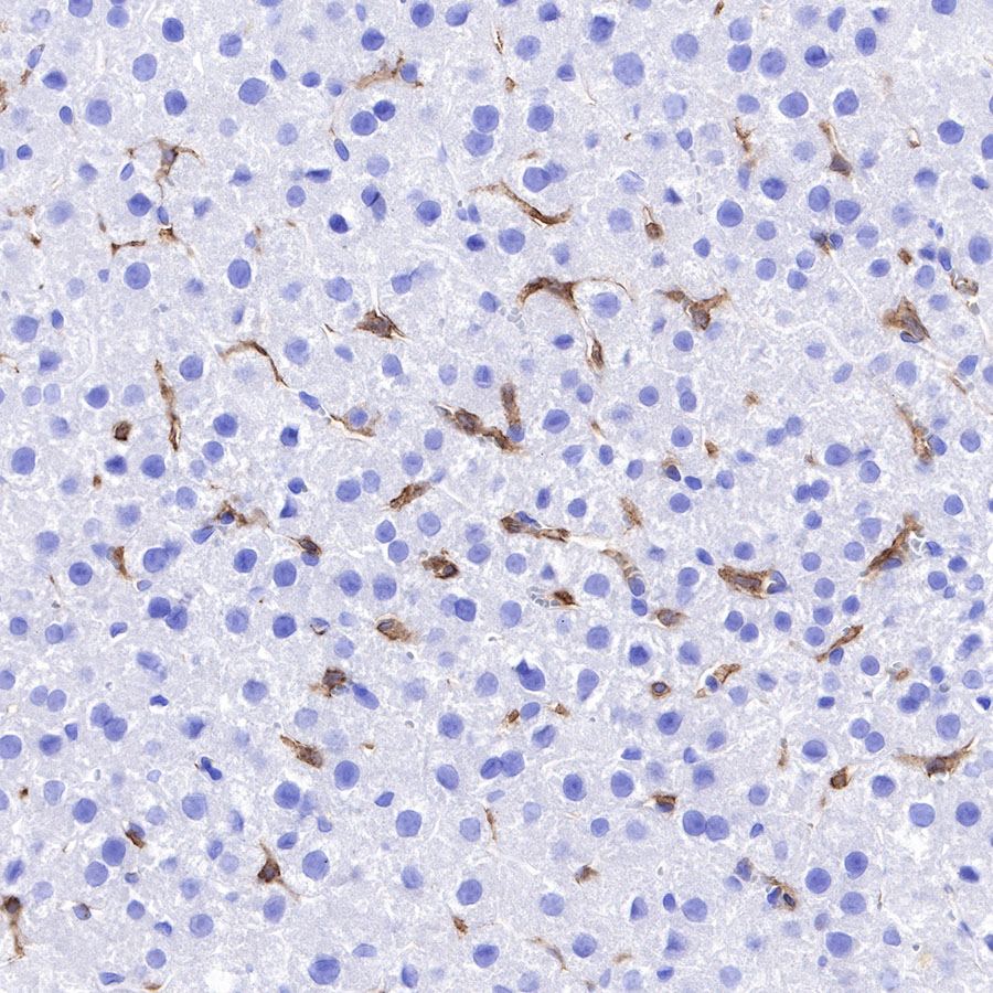
IHC shows positive staining in paraffin-embedded rat liver. Anti-F4/80 antibody was used at 1/500 dilution, followed by a HRP Polymer for Mouse & Rabbit IgG (ready to use). Counterstained with hematoxylin. Heat mediated antigen retrieval with Tris/EDTA buffer pH9.0 was performed before commencing with IHC staining protocol.