
Rabbit anti-HDAC1 Recombinant Monoclonal Antibody(310-72)别名宿主反应种属应用免疫原形式浓度纯化方法类型克隆号储存/保存方法存储溶液背景说明细胞定位UniProt
| 概述 | |
| 别名 |
Histone deacetylase 1; HD1; Protein deacetylase HDAC1
|
| 宿主 |
Rabbit
|
| 反应种属 |
Human, Mouse, Rat
|
| 应用 |
WB: 1:5000, IHC-P: 1:200
|
| 免疫原 |
Synthetic peptide
|
| 性能 | |
| 形式 |
Liquid
|
| 浓度 |
0.05 mg/mL
|
| 纯化方法 |
Protein A affinity column
|
| 类型 |
Monoclonal Antibody
|
| 克隆号 |
310-72
|
| 储存/保存方法 |
Store at -20℃ for one year.
|
| 存储溶液 |
PBS, 40% Glycerol, 0.05% BSA, 0.03% Proclin 300
|
| 靶标 | |
| 背景说明 |
HDAC family has four subclasses including I, II, III and IV. Histone deacetylase 1 (HDAC1) is a unique member of the classes I HDACs that has been shown to be involved in gene transcription, transcriptional regulation, cell cycle progression and developmental events by controlling both enzyme activity and epigenetics of key proteins [PMID: 16289629]. HDAC1 is the most abundant member of the class I HDACs in pulmonary endothelial cells [PMID: 34264338], regulating the enzymatic activity and epigenetics of key proteins to adapt to external stimuli. It can efficiently decrotonylate this relatively less abundant histone modification [PMID: 29317660].
|
| 细胞定位 |
Nucleus
|
| UniProt |
Q13547
|
实验结果图
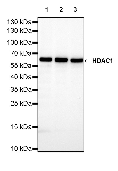
WB result of HDAC1 Rabbit mAb Primary antibody: HDAC1 Rabbit mAb at 1/5000 dilution Lane 1: HeLa whole cell lysate 20 µg Lane 2: Jurkat whole cell lysate 20 µg Lane 3: K-562 whole cell lysate 20 µg Secondary antibody: #JP20040 at 1/10000 dilution Predicted MW: 55kDa Observed MW: 62kDa Exposure time: 20s
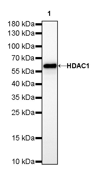
WB result of HDAC1 Rabbit mAb Primary antibody: HDAC1 Rabbit mAb at 1/5000 dilution Lane 1: NIH/3T3 whole cell lysate 20 µg Secondary antibody: #JP20040 at 1/10000 dilution Predicted MW: 55kDa Observed MW: 62kDa Exposure time: 20s
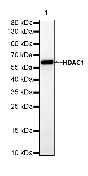
WB result of HDAC1 Rabbit mAb Primary antibody: HDAC1 Rabbit mAb at 1/5000 dilution Lane 1: C6 whole cell lysate 20 µg Secondary antibody: #JP20040 at 1/10000 dilution Predicted MW: 55kDa Observed MW: 62kDa Exposure time: 20s
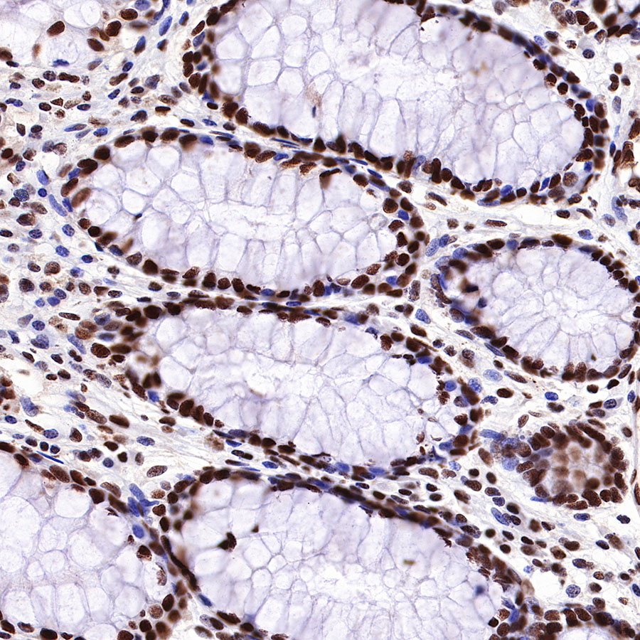
IHC shows positive staining in paraffin-embedded human colon. Anti-HDAC1 antibody was used at 1/200 dilution, followed by a HRP Polymer for Mouse & Rabbit IgG (ready to use). Counterstained with hematoxylin. Heat mediated antigen retrieval with Tris/EDTA buffer pH9.0 was performed before commencing with IHC staining protocol.
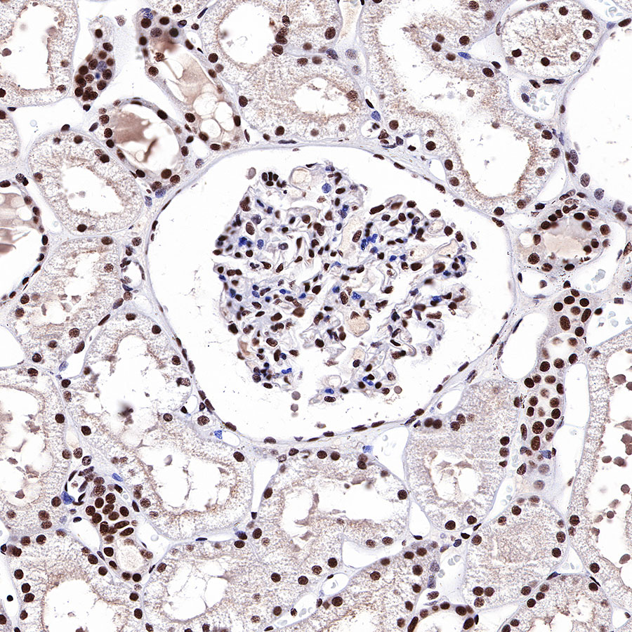
IHC shows positive staining in paraffin-embedded human kidney. Anti-HDAC1 antibody was used at 1/200 dilution, followed by a HRP Polymer for Mouse & Rabbit IgG (ready to use). Counterstained with hematoxylin. Heat mediated antigen retrieval with Tris/EDTA buffer pH9.0 was performed before commencing with IHC staining protocol.

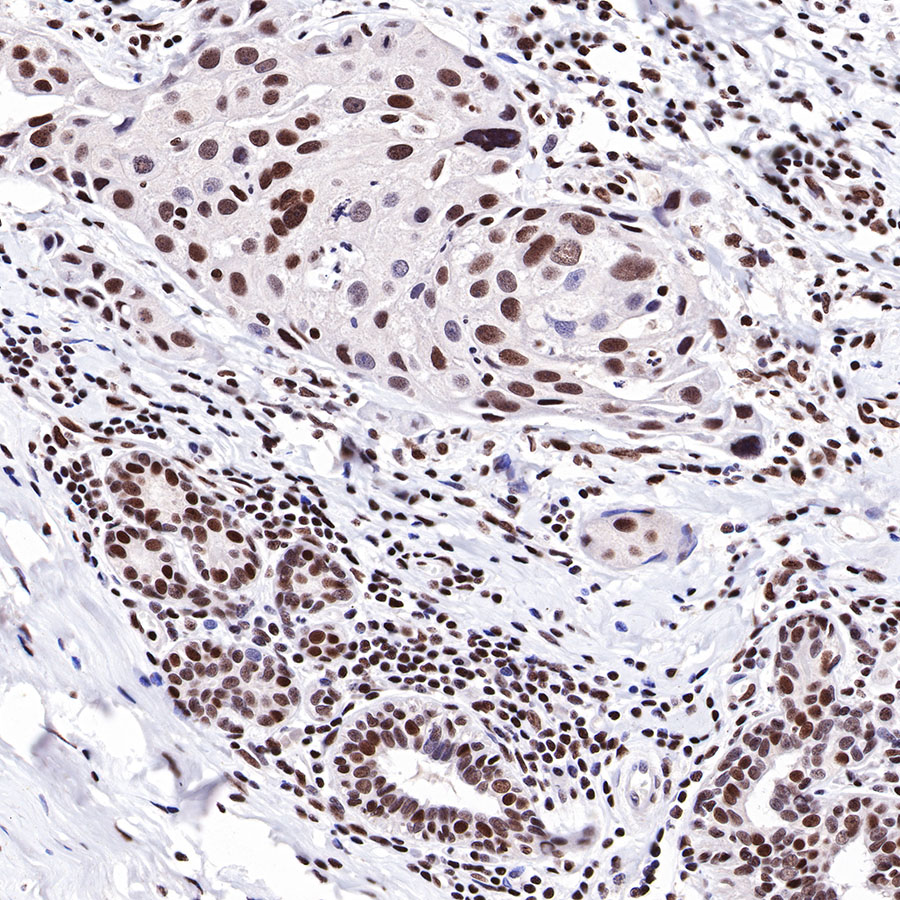
IHC shows positive staining in paraffin-embedded human colon cancer. Anti-HDAC1 antibody was used at 1/200 dilution, followed by a HRP Polymer for Mouse & Rabbit IgG (ready to use). Counterstained with hematoxylin. Heat mediated antigen retrieval with Tris/EDTA buffer pH9.0 was performed before commencing with IHC staining protocol.
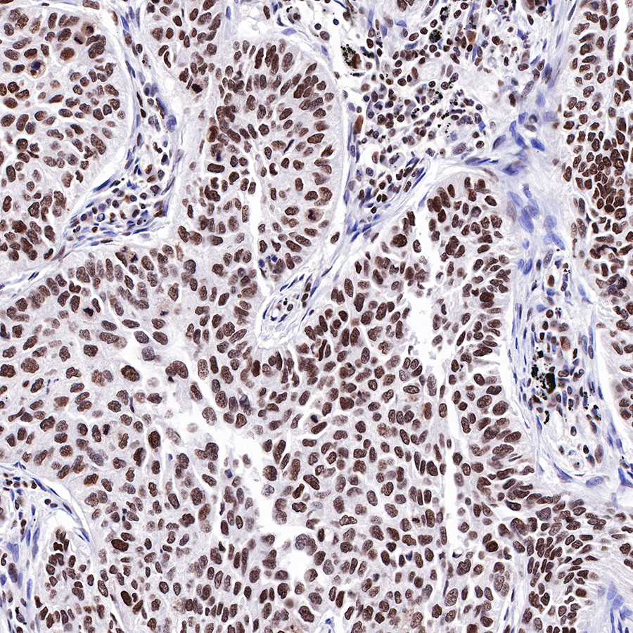
IHC shows positive staining in paraffin-embedded human breast cancer. Anti-HDAC1 antibody was used at 1/200 dilution, followed by a HRP Polymer for Mouse & Rabbit IgG (ready to use). Counterstained with hematoxylin. Heat mediated antigen retrieval with Tris/EDTA buffer pH9.0 was performed before commencing with IHC staining protocol.
IHC shows positive staining in paraffin-embedded human lung squamous cell carcinoma. Anti-HDAC1 antibody was used at 1/200 dilution, followed by a HRP Polymer for Mouse & Rabbit IgG (ready to use). Counterstained with hematoxylin. Heat mediated antigen retrieval with Tris/EDTA buffer pH9.0 was performed before commencing with IHC staining protocol.