
Rabbit anti-MEK1 Recombinant Monoclonal Antibody(308-32)别名宿主反应种属应用免疫原形式浓度纯化方法类型克隆号储存/保存方法存储溶液背景说明细胞定位UniProt
| 概述 | |
| 别名 |
Dual specificity mitogen-activated protein kinase kinase 1; MAP kinase kinase 1; MAPKK 1; MKK1; ERK activator kinase 1; MAPK/ERK kinase 1
|
| 宿主 |
Rabbit
|
| 反应种属 |
Human
|
| 应用 |
WB: 1:1000, IP: 1:50, IHC-P: 1:500, ICC: 1:1000
|
| 免疫原 |
Synthetic peptide
|
| 性能 | |
| 形式 |
Liquid
|
| 浓度 |
0.5 mg/mL
|
| 纯化方法 |
Protein A affinity column
|
| 类型 |
Monoclonal Antibody
|
| 克隆号 |
308-32
|
| 储存/保存方法 |
Store at -20℃ for one year.
|
| 存储溶液 |
PBS, 40% Glycerol, 0.05% BSA, 0.03% Proclin 300
|
| 靶标 | |
| 背景说明 |
Mitogen-activated protein kinase kinases 1 and 2 (MEK1/2) are the crucial part of the RAS-RAF-MEK-ERK pathway (or ERK pathway), which is involved in the regulation of various cellular processes including proliferation, survival, and differentiation et al. Targeting MEK has become an important strategy for cancer therapy, and 4 MEK inhibitors (MEKis) have been approved by FDA to date [PMID: 33774345].
|
| 细胞定位 |
Cytoplasm, cytoskeleton, Nucleus, Membrane
|
| UniProt |
Q02750
|
实验结果图
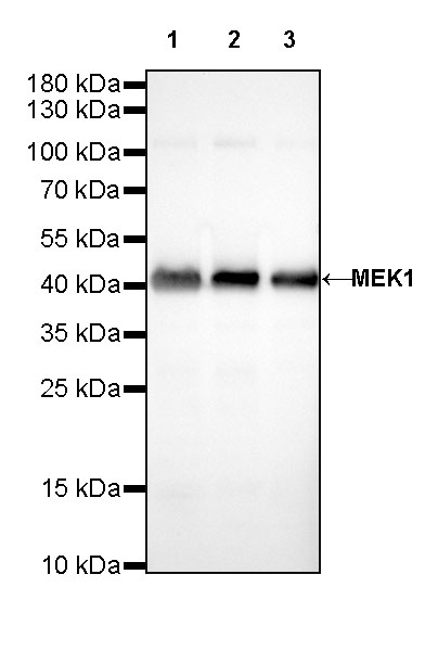
WB result of MEK1 Rabbit mAb Primary antibody: MEK1 Rabbit mAb at 1/1000 dilution Lane 1: HeLa whole cell lysate 20ug Lane 2: Jurkat whole cell lysate 20ug Lane 3: A431 whole cell lysate 20ug Secondary antibody: #JP20040 at 1/10000 dilution Predicted MW: 43 kDa Observed MW: 43 kDa Exposure time: 30s
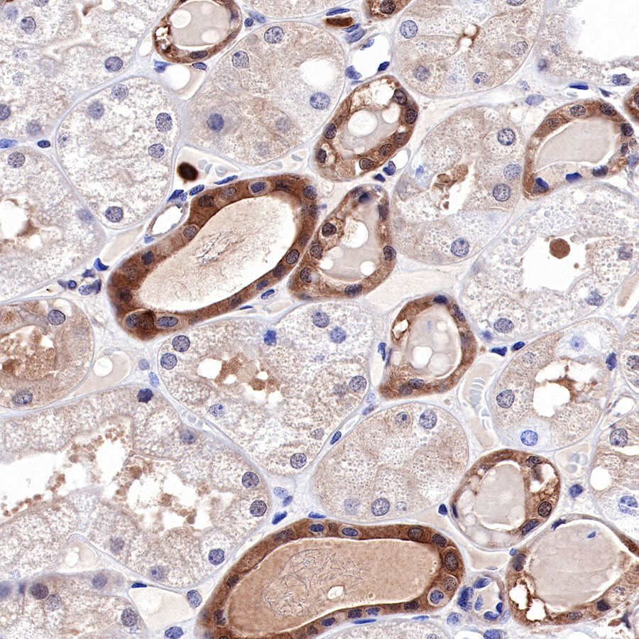
IHC shows positive staining in paraffin-embedded human kidney. Anti-MEK1 antibody was used at 1/500 dilution, followed by a HRP Polymer for Mouse & Rabbit IgG (ready to use). Counterstained with hematoxylin. Heat mediated antigen retrieval with Tris/EDTA buffer pH9.0 was performed before commencing with IHC staining protocol.
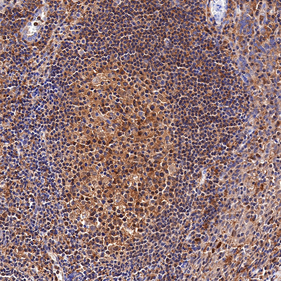
IHC shows positive staining in paraffin-embedded human tonsil. Anti-MEK1 antibody was used at 1/500 dilution, followed by a HRP Polymer for Mouse & Rabbit IgG (ready to use). Counterstained with hematoxylin. Heat mediated antigen retrieval with Tris/EDTA buffer pH9.0 was performed before commencing with IHC staining protocol.
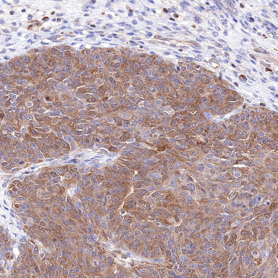
IHC shows positive staining in paraffin-embedded human ovarian carcinoma. Anti-MEK1 antibody was used at 1/500 dilution, followed by a HRP Polymer for Mouse & Rabbit IgG (ready to use). Counterstained with hematoxylin. Heat mediated antigen retrieval with Tris/EDTA buffer pH9.0 was performed before commencing with IHC staining protocol.
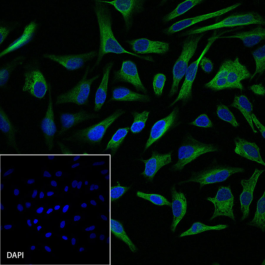
ICC shows positive staining in HeLa cells. Anti-MEK1 antibody was used at 1/1000 dilution and incubated overnight at 4°C. Goat polyclonal Antibody to Rabbit IgG – H&L (Alexa Fluor® 488) was used as secondary antibody at 1/1000 dilution. The cells were fixed with 4% PFA and permeabilized with 0.1% PBS-Triton X-100. Nuclei were counterstained with DAPI.
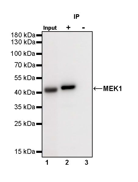
MEK1 Rabbit mAb at 1/50 dilution (1 µg) immunoprecipitating MEK1 in 0.4 mg A431 whole cell lysate. Western blot was performed on the immunoprecipitate using MEK1 Rabbit mAb at 1/1000 dilution. Secondary antibody (HRP) for IP was used at 1/400 dilution. Lane 1: A431 whole cell lysate 20 µg (Input) Lane 2: MEK1 Rabbit mAb IP in A431 whole cell lysate Lane 3: Rabbit monoclonal IgG IP in A431 whole cell lysate Predicted MW: 43 kDa Observed MW: 43 kDa Exposure time: 40 s