
Rabbit anti-Lamin B1 Recombinant Monoclonal Antibody(307-108)别名宿主反应种属应用免疫原形式浓度纯化方法类型克隆号储存/保存方法存储溶液背景说明细胞定位UniProt
| 概述 | |
| 别名 |
LMNB1; Lamin-B1
|
| 宿主 |
Rabbit
|
| 反应种属 |
Human, Mouse, Rat
|
| 应用 |
WB: 1:1000, IHC-P: 1:2000
|
| 免疫原 |
Synthetic peptide
|
| 性能 | |
| 形式 |
Liquid
|
| 浓度 |
0.5 mg/mL
|
| 纯化方法 |
Protein A affinity column
|
| 类型 |
Monoclonal Antibody
|
| 克隆号 |
307-108
|
| 储存/保存方法 |
Store at -20℃ for one year.
|
| 存储溶液 |
PBS, 40% Glycerol, 0.05% BSA, 0.03% Proclin 300
|
| 靶标 | |
| 背景说明 |
Lamin-B1 is a protein that in humans is encoded by the LMNB1 gene. It is one of the essential members of the nuclear lamina protein family. Its main function is to maintain the integrity of nuclear skeleton, as well as to participate in the cell proliferation and aging by affecting the chromosome distribution. gene expression, and DNA damage repair. The abnormal expression of lamin B1 is related to certain diseases, including neurological diseases [e.g. neural tube defects (NDTs), adult-onset autosomal dominant leukodystrophy (ADLD)] and tumors (e.g. pancreatic cancer). It is also a potential tumor marker as well as drug target [PMID: 30499270].
|
| 细胞定位 |
Nucleus lamina
|
| UniProt |
P20700
|
实验结果图
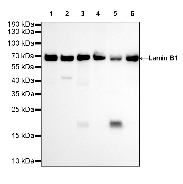
WB result of Lamin B1 Rabbit mAb Primary antibody: Lamin B1 Rabbit mAb at 1/1000 dilution Lane 1: HeLa whole cell lysate 20 ug Lane 2: Ramos whole cell lysate 20 ug Lane 3: MCF7 whole cell lysate 20 ug Lane 4: Jurkat whole cell lysate 20 ug Lane 5: MOLT-4 whole cell lysate 20 ug Lane 6: Caco-2 whole cell lysate 20 ug Secondary antibody: Goat Anti-Rabbit IgG, (H+L), HRP conjugated at 1/10000 dilution Predicted MW: 68 kDa Observed MW: 68 kDa Exposure time: 60 s
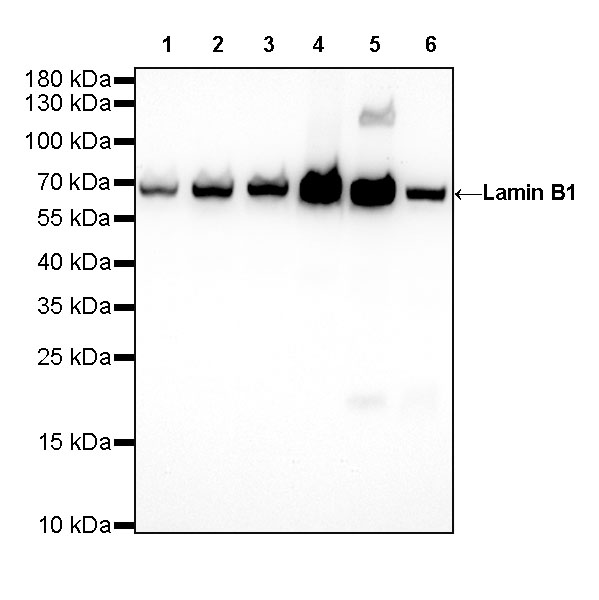
WB result of Lamin B1 Rabbit mAb Primary antibody: Lamin B1 Rabbit mAb at 1/1000 dilution Lane 1: NIH/3T3 whole cell lysate 20 ug Lane 2: RAW 264.7 whole cell lysate 20 ug Lane 3: mouse brain lysate 20 ug Lane 4: mouse heart lysate 20 ug Lane 5: mouse kidney lysate 20 ug Lane 6: mouse spleen lysate 20 ug Secondary antibody: Goat Anti-Rabbit IgG, (H+L), HRP conjugated at 1/10000 dilution Predicted MW: 68 kDa Observed MW: 68 kDa Exposure time: 20 s
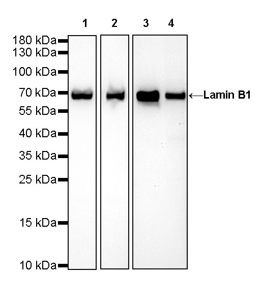
WB result of Lamin B1 Rabbit mAb Primary antibody: Lamin B1 Rabbit mAb at 1/1000 dilution Lane 1: PC-12 whole cell lysate 20 ug Lane 2: rat brain lysate 20 ug Lane 3: rat heart lysate 20 ug Lane 4: rat kidney lysate 20 ug Secondary antibody: Goat Anti-Rabbit IgG, (H+L), HRP conjugated at 1/10000 dilution Predicted MW: 68 kDa Observed MW: 68 kDa Exposure time: 20 s
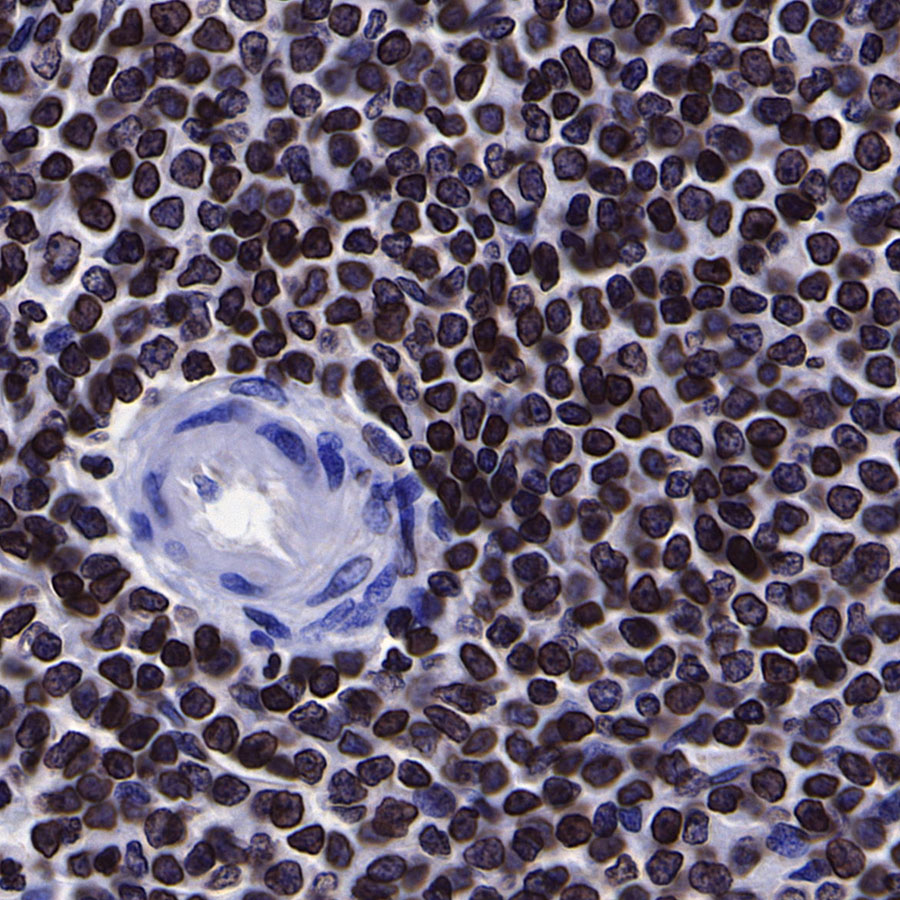
IHC shows positive staining in paraffin-embedded human spleen. Anti-Lamin B1 antibody was used at 1/2000 dilution, followed by a Goat Anti-Rabbit IgG H&L (HRP) ready to use. Counterstained with hematoxylin. Heat mediated antigen retrieval with Tris/EDTA buffer pH9.0 was performed before commencing with IHC staining protocol.
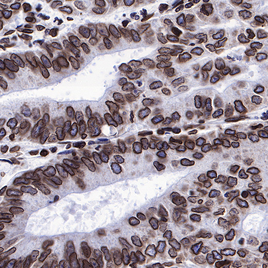
IHC shows positive staining in paraffin-embedded human stomach. Anti-Lamin B1 antibody was used at 1/2000 dilution, followed by a Goat Anti-Rabbit IgG H&L (HRP) ready to use. Counterstained with hematoxylin. Heat mediated antigen retrieval with Tris/EDTA buffer pH9.0 was performed before commencing with IHC staining protocol.
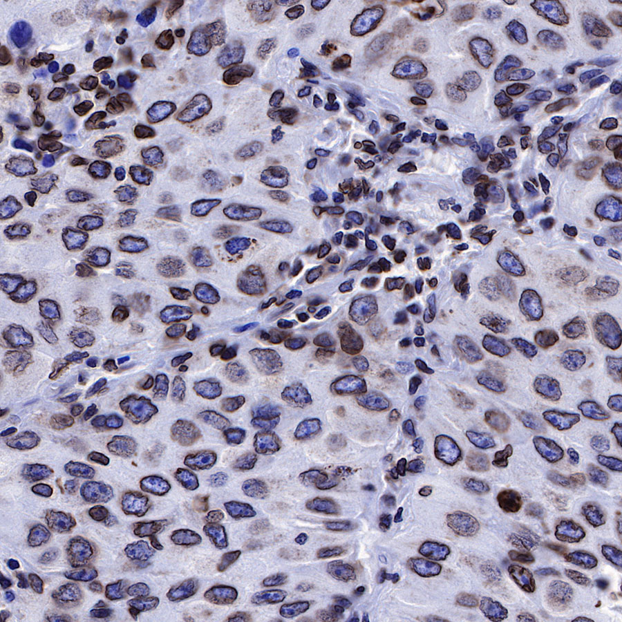
IHC shows positive staining in paraffin-embedded human endometrial cancer. Anti-Lamin B1 antibody was used at 1/2000 dilution, followed by a Goat Anti-Rabbit IgG H&L (HRP) ready to use. Counterstained with hematoxylin. Heat mediated antigen retrieval with Tris/EDTA buffer pH9.0 was performed before commencing with IHC staining protocol.
IHC shows positive staining in paraffin-embedded human lung cancer. Anti-Lamin B1 antibody was used at 1/2000 dilution, followed by a Goat Anti-Rabbit IgG H&L (HRP) ready to use. Counterstained with hematoxylin. Heat mediated antigen retrieval with Tris/EDTA buffer pH9.0 was performed before commencing with IHC staining protocol.
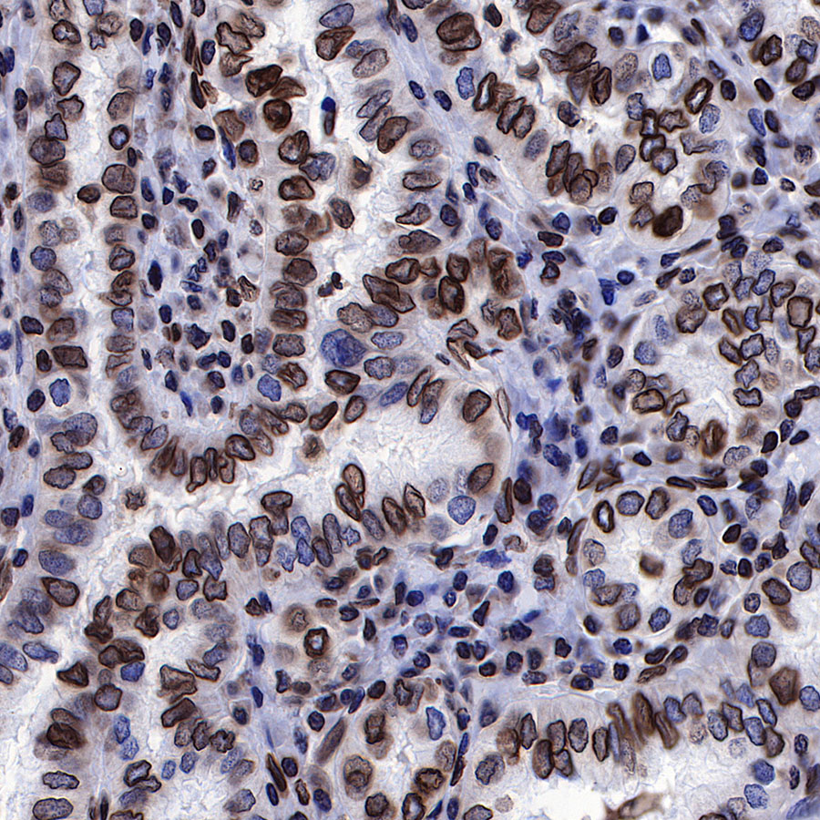
IHC shows positive staining in paraffin-embedded human thyroid cancer. Anti-Lamin B1 antibody was used at 1/2000 dilution, followed by a Goat Anti-Rabbit IgG H&L (HRP) ready to use. Counterstained with hematoxylin. Heat mediated antigen retrieval with Tris/EDTA buffer pH9.0 was performed before commencing with IHC staining protocol.
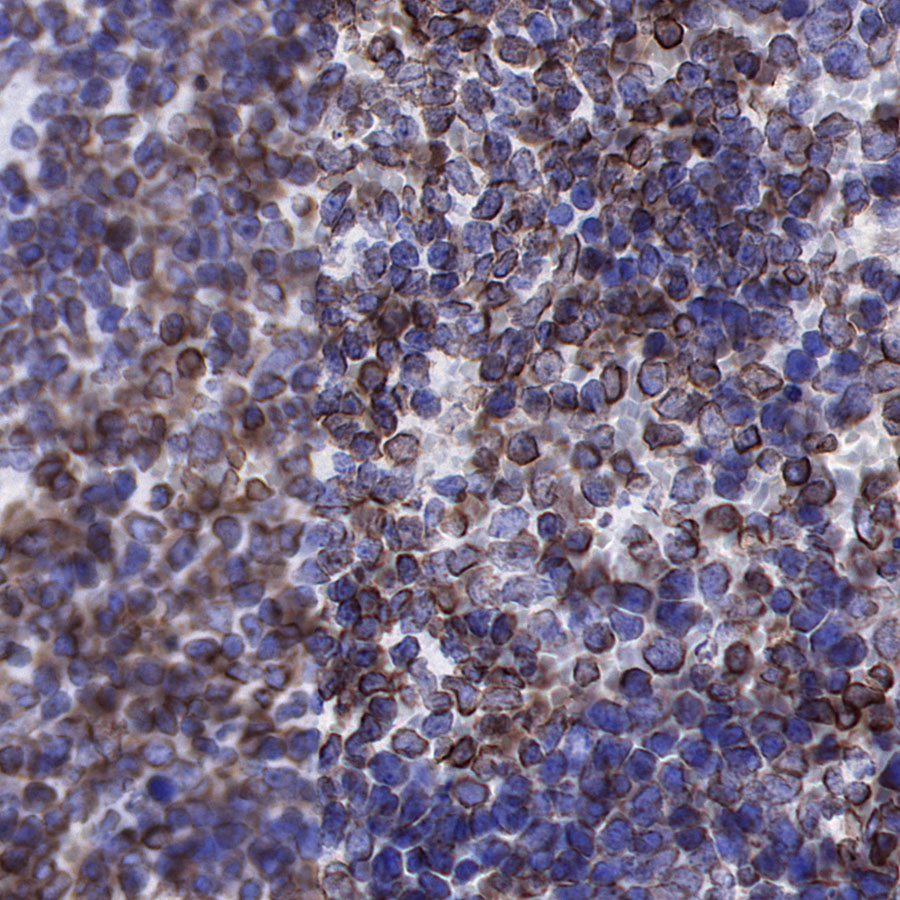
IHC shows positive staining in paraffin-embedded mouse spleen. Anti-Lamin B1 antibody was used at 1/2000 dilution, followed by a Goat Anti-Rabbit IgG H&L (HRP) ready to use. Counterstained with hematoxylin. Heat mediated antigen retrieval with Tris/EDTA buffer pH9.0 was performed before commencing with IHC staining protocol.
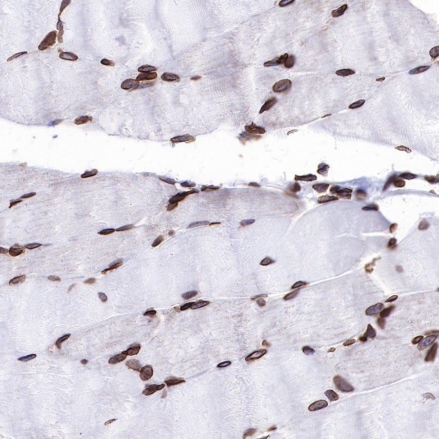
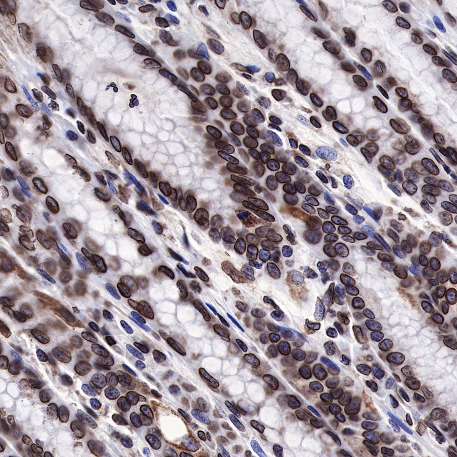
IHC shows positive staining in paraffin-embedded mouse skeletal muscle. Anti-Lamin B1 antibody was used at 1/2000 dilution, followed by a Goat Anti-Rabbit IgG H&L (HRP) ready to use. Counterstained with hematoxylin. Heat mediated antigen retrieval with Tris/EDTA buffer pH9.0 was performed before commencing with IHC staining protocol.
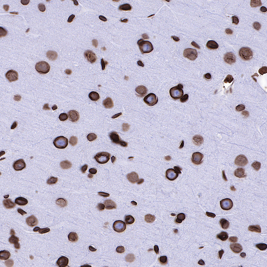
IHC shows positive staining in paraffin-embedded rat cerebral cortex. Anti-Lamin B1 antibody was used at 1/2000 dilution, followed by a Goat Anti-Rabbit IgG H&L (HRP) ready to use. Counterstained with hematoxylin. Heat mediated antigen retrieval with Tris/EDTA buffer pH9.0 was performed before commencing with IHC staining protocol.
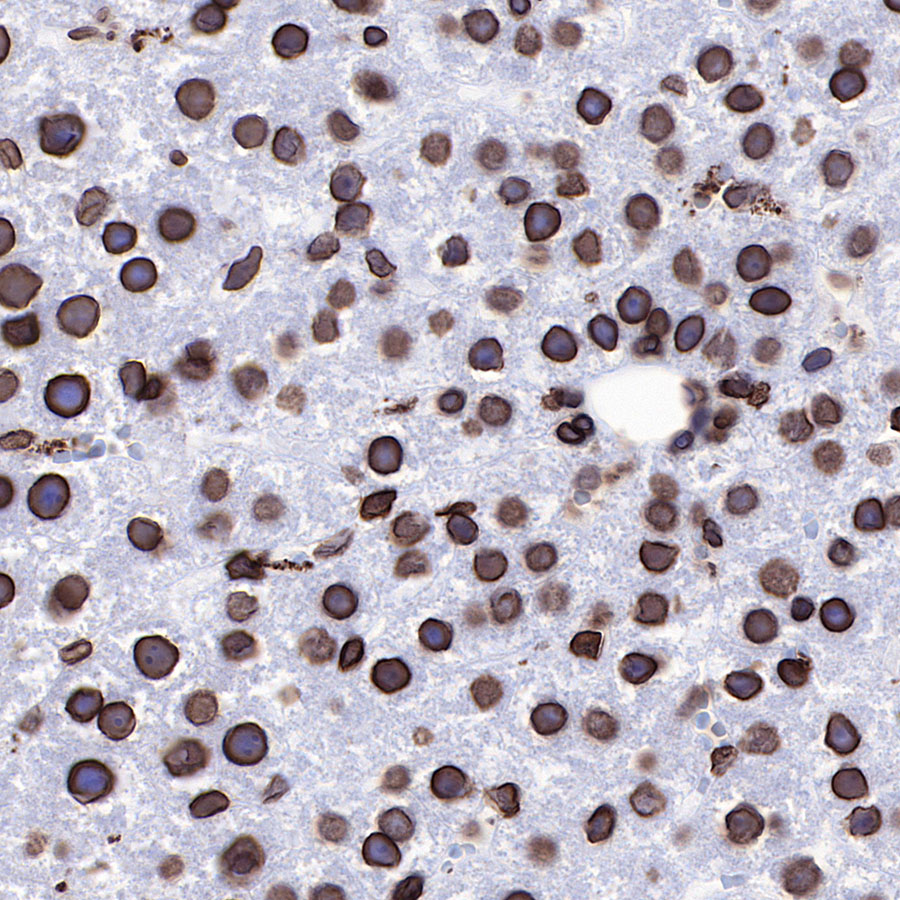
IHC shows positive staining in paraffin-embedded rat liver. Anti-Lamin B1 antibody was used at 1/2000 dilution, followed by a Goat Anti-Rabbit IgG H&L (HRP) ready to use. Counterstained with hematoxylin. Heat mediated antigen retrieval with Tris/EDTA buffer pH9.0 was performed before commencing with IHC staining protocol.