
Rabbit anti-GFAP Recombinant Monoclonal Antibody(306-10)描述别名宿主特异性反应种属预测反应种属应用免疫原形式浓度纯化方法类型克隆号储存/保存方法存储溶液背景说明组织特异性翻译后修饰细胞定位UniProt
| 概述 | |
| 描述 |
This gene encodes one of the major intermediate filament proteins of mature astrocytes. It is used as a marker to distinguish astrocytes from other glial cells during development. Mutations in this gene cause Alexander disease, a rare disorder of astrocytes in the central nervous system. An additional transcript variant has been described, but its full length sequence has not been determined.
|
| 别名 |
GFAP抗体;Glial fibrillary acidic protein
|
| 宿主 |
Rabbit
|
| 特异性 |
GFAP Antibody detects endogenous levels of total GFAP.
|
| 反应种属 |
Human, Mouse, Rat
|
| 预测反应种属 |
Horse;Bovine;
|
| 应用 |
WB: 1:500, IHC-P: 1:1000
|
| 免疫原 |
Synthetic Peptide
|
| 性能 | |
| 形式 |
Liquid
|
| 浓度 |
0.25 mg/mL
|
| 纯化方法 |
Protein A affinity column
|
| 类型 |
Monoclonal Antibody
|
| 克隆号 |
306-10
|
| 储存/保存方法 |
Store at -20℃ for one year.
|
| 存储溶液 |
PBS, 40% Glycerol, 0.05% BSA, 0.03% Proclin 300
|
| 靶标 | |
| 背景说明 |
Glial fibrillary acidic protein (GFAP) is the hallmark intermediate filament (IF; also known as nanofilament) protein in astrocytes, a main type of glial cells in the central nervous system (CNS). Astrocytes have a range of control and homeostatic functions in health and disease. Astrocytes assume a reactive phenotype in acute CNS trauma, ischemia, and in neurodegenerative diseases. This coincides with an upregulation and rearrangement of the IFs, which form a highly complex system composed of GFAP (10 isoforms), vimentin, synemin, and nestin [PMID: 25726916].
|
| 组织特异性 |
Expressed in cells lacking fibronectin.
|
| 翻译后修饰 |
Phosphorylated by PKN1.
|
| 细胞定位 |
Cytoplasm
|
| UniProt |
P14136
|
实验结果图
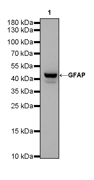
WB result of GFAP Rabbit mAb Primary antibody: GFAP Rabbit mAb at 1/1000 dilution Lane 1: mouse brain lysate 20 µg Secondary antibody: Goat Anti-Rabbit IgG, (H+L), HRP conjugated at 1/10000 dilution Predicted MW: 50kDa Observed MW: 42kDa Exposure time: 30s
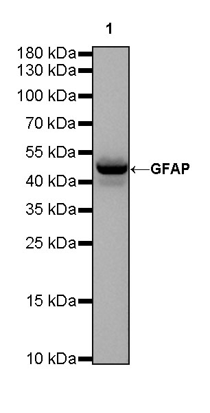
WB result of GFAP Rabbit mAb Primary antibody: GFAP Rabbit mAb at 1/1000 dilution Lane 1: rat brain lysate 20 µg Secondary antibody: Goat Anti-Rabbit IgG, (H+L), HRP conjugated at 1/10000 dilution Predicted MW: 50kDa Observed MW: 50kDa Exposure time: 30s
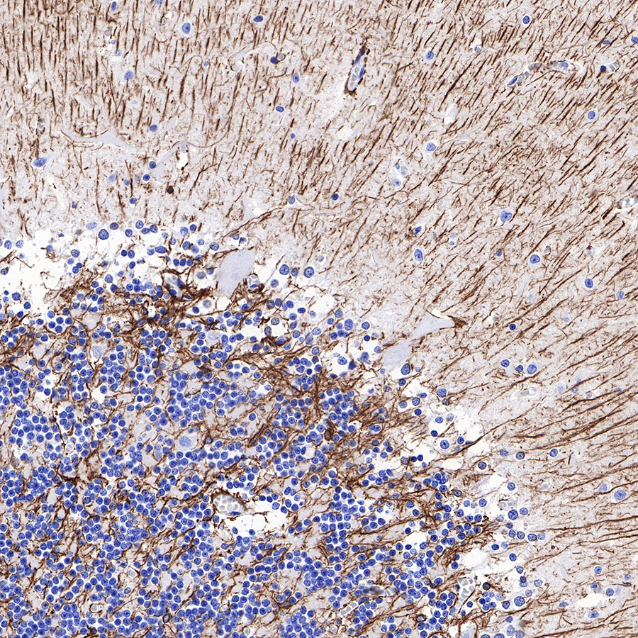
IHC shows positive staining in paraffin-embedded human cerebellum. Anti-GFAP antibody was used at 1/1000 dilution, followed by a HRP Polymer for Mouse & Rabbit IgG (ready to use). Counterstained with hematoxylin. Heat mediated antigen retrieval with Tris/EDTA buffer pH9.0 was performed before commencing with IHC staining protocol.
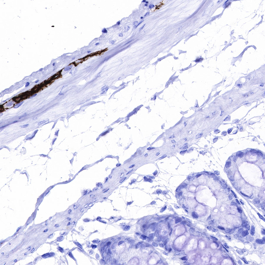
IHC shows positive staining in paraffin-embedded rat colon. Anti-GFAP antibody was used at 1/1000 dilution, followed by a HRP Polymer for Mouse & Rabbit IgG (ready to use). Counterstained with hematoxylin. Heat mediated antigen retrieval with Tris/EDTA buffer pH9.0 was performed before commencing with IHC staining protocol.
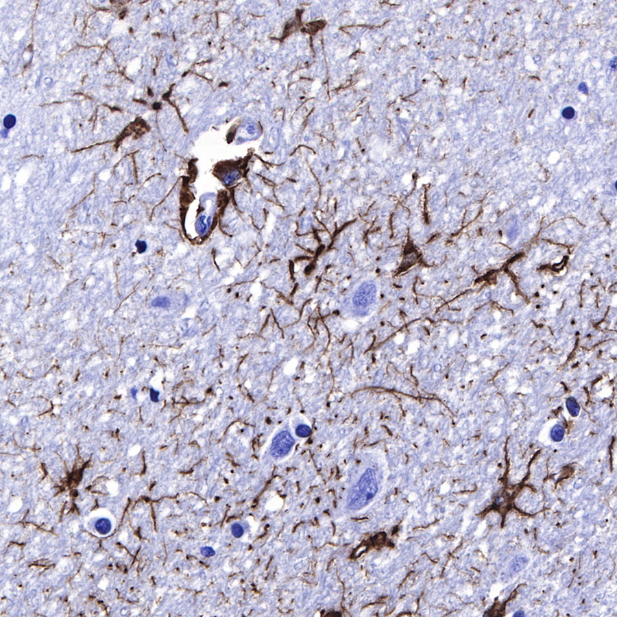
IHC shows positive staining in paraffin-embedded human cerebral cortex. Anti-GFAP antibody was used at 1/1000 dilution, followed by a HRP Polymer for Mouse & Rabbit IgG (ready to use). Counterstained with hematoxylin. Heat mediated antigen retrieval with Tris/EDTA buffer pH9.0 was performed before commencing with IHC staining protocol.
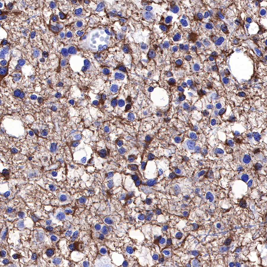
IHC shows positive staining in paraffin-embedded human glioma. Anti-GFAP antibody was used at 1/1000 dilution, followed by a HRP Polymer for Mouse & Rabbit IgG (ready to use). Counterstained with hematoxylin. Heat mediated antigen retrieval with Tris/EDTA buffer pH9.0 was performed before commencing with IHC staining protocol.
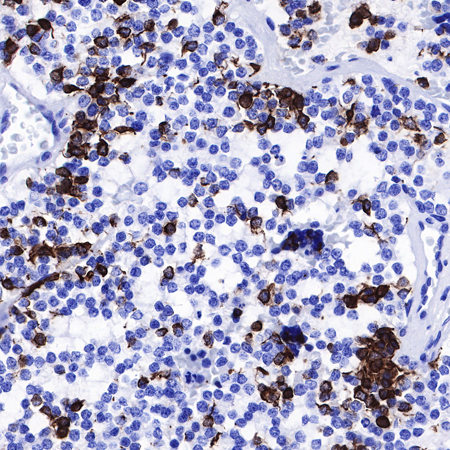
IHC shows positive staining in paraffin-embedded human astrocytoma. Anti-GFAP antibody was used at 1/1000 dilution, followed by a HRP Polymer for Mouse & Rabbit IgG (ready to use). Counterstained with hematoxylin. Heat mediated antigen retrieval with Tris/EDTA buffer pH9.0 was performed before commencing with IHC staining protocol.
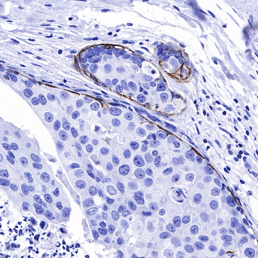
IHC shows positive staining in paraffin-embedded human breast cancer. Anti-GFAP antibody was used at 1/1000 dilution, followed by a HRP Polymer for Mouse & Rabbit IgG (ready to use). Counterstained with hematoxylin. Heat mediated antigen retrieval with Tris/EDTA buffer pH9.0 was performed before commencing with IHC staining protocol.
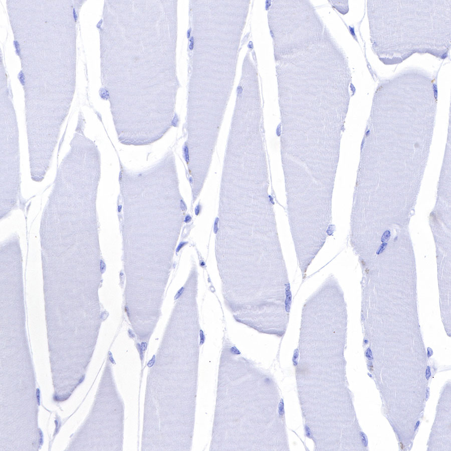
Negative control: IHC shows negative staining in paraffin-embedded human skeletal muscle. Anti-GFAP antibody was used at 1/1000 dilution, followed by a HRP Polymer for Mouse & Rabbit IgG (ready to use). Counterstained with hematoxylin. Heat mediated antigen retrieval with Tris/EDTA buffer pH9.0 was performed before commencing with IHC staining protocol.
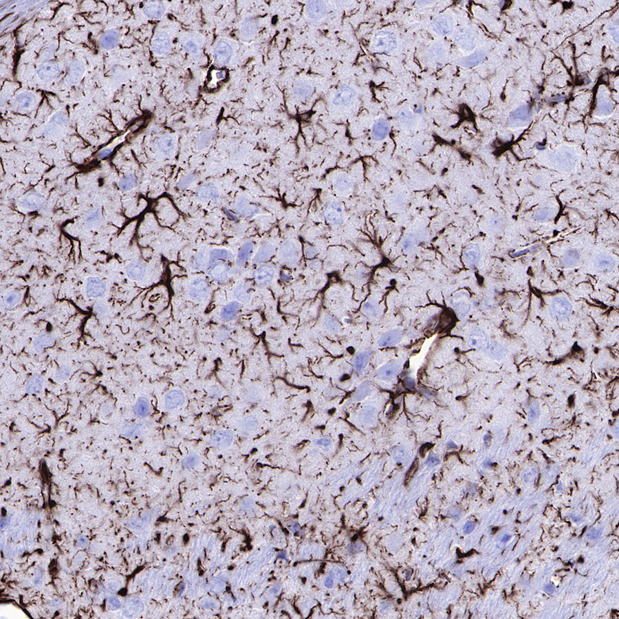
IHC shows positive staining in paraffin-embedded mouse cerebral cortex. Anti-GFAP antibody was used at 1/1000 dilution, followed by a HRP Polymer for Mouse & Rabbit IgG (ready to use). Counterstained with hematoxylin. Heat mediated antigen retrieval with Tris/EDTA buffer pH9.0 was performed before commencing with IHC staining protocol.
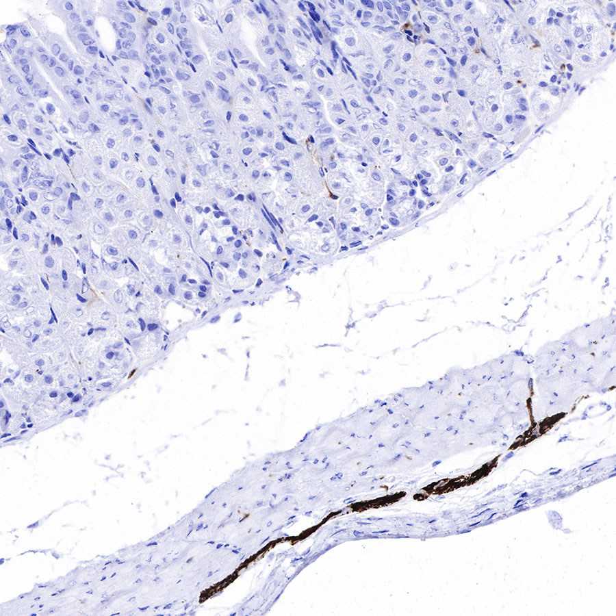
IHC shows positive staining in paraffin-embedded mouse stomach. Anti-GFAP antibody was used at 1/1000 dilution, followed by a HRP Polymer for Mouse & Rabbit IgG (ready to use). Counterstained with hematoxylin. Heat mediated antigen retrieval with Tris/EDTA buffer pH9.0 was performed before commencing with IHC staining protocol.
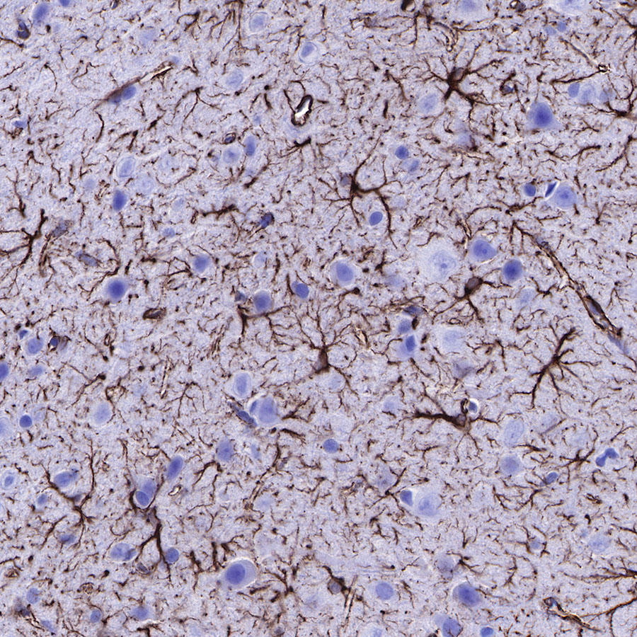
IHC shows positive staining in paraffin-embedded rat cerebral cortex. Anti-GFAP antibody was used at 1/1000 dilution, followed by a HRP Polymer for Mouse & Rabbit IgG (ready to use). Counterstained with hematoxylin. Heat mediated antigen retrieval with Tris/EDTA buffer pH9.0 was performed before commencing with IHC staining protocol.