
Rabbit anti-p16 Recombinant Monoclonal Antibody(S-303-1)描述别名宿主特异性反应种属应用免疫原形式浓度纯化方法类型克隆号储存/保存方法存储溶液背景说明组织特异性翻译后修饰细胞定位UniProt
| 概述 | |
| 描述 |
p16-INK4A a cell-cycle regulatory protein that Interacts with CDK4 and CDK6, inhibiting their ability to interact with cyclins D. Inhibits the phosphorylation of the retinoblastoma protein by CDK4 or CDK6. Four alternatively spliced isoforms have been reported.
|
| 别名 |
p16 INK抗体;Cyclin-dependent kinase inhibitor 2A; Cyclin-dependent kinase 4 inhibitor A (CDK4I); Multiple tumor suppressor 1 (MTS-1); p16-INK4a (p16-INK4; p16INK4A); CDKN2A; CDKN2; MTS1
|
| 宿主 |
Rabbit
|
| 特异性 |
p16 INK antibody detects endogenous levels of total p16 INK.
|
| 反应种属 |
Human
|
| 应用 |
WB: 1:1000, IP: 1:50, ICC: 1:100, FC(Intra): 1:500
|
| 免疫原 |
Recombinant protein
|
| 性能 | |
| 形式 |
Liquid
|
| 浓度 |
0.5 mg/mL
|
| 纯化方法 |
Protein A affinity column
|
| 类型 |
Monoclonal Antibody
|
| 克隆号 |
S-303-1
|
| 储存/保存方法 |
Store at -20℃ for one year.
|
| 存储溶液 |
PBS, 40% Glycerol, 0.05% BSA, 0.03% Proclin 300
|
| 靶标 | |
| 背景说明 |
p16 (also known as p16INK4a, cyclin-dependent kinase inhibitor 2A, CDKN2A, multiple tumor suppressor 1 and numerous other synonyms), is a protein that slows cell division by slowing the progression of the cell cycle from the G1 phase to the S phase, thereby acting as a tumor suppressor. p16 can be used as a biomarker to improve the histological diagnostic accuracy of grade 3 cervical intraepithelial neoplasia (CIN). p16 is also implicated in the prevention of melanoma, oropharyngeal squamous cell carcinoma, cervical cancer, vulvar cancer and esophageal cancer.
|
| 组织特异性 |
Widely expressed but not detected in brain or skeletal muscle. Isoform 3 is pancreas-specific.
|
| 翻译后修饰 |
Phosphorylation seems to increase interaction with CDK4.
|
| 细胞定位 |
Cytoplasm, Nucleus
|
| UniProt |
P42771
|
实验结果图
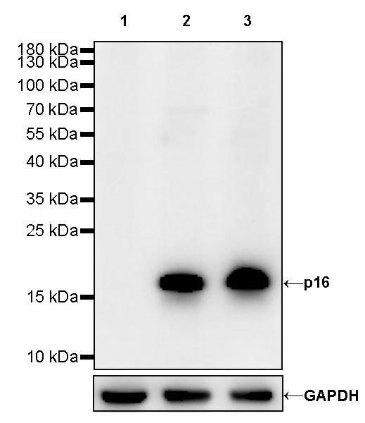
WB result of p16 Rabbit mAb Primary antibody: p16 Rabbit mAb at 1/1000 dilution Lane 1: MCF7 whole cell lysate 20 ug Lane 2: HeLa whole cell lysate 20 ug Lane 3: HEK-293 whole cell lysate 20 ug Negative control: MCF7 whole cell lysate Secondary antibody: Goat Anti-Rabbit IgG, (H+L), HRP conjugated at 1/10000 dilution Predicted MW: 16 kDa Observed MW: 16 kDa Exposure time: 30 s (This blot was developed with high sensitivity substrate)

p16 Rabbit mAb at 1/50 dilution (1 ug) immunoprecipitating p16 in 0.4 mg HeLa whole cell lysate. Western blot was performed on the immunoprecipitate using p16 Rabbit mAb at 1/1000 dilution. Secondary antibody (HRP) for IP was used at 1/400 dilution. Lane 1: HeLa whole cell lysate 20 ug (Input) Lane 2: p16 Rabbit mAb IP in HeLa whole cell lysate Lane 3: Rabbit monoclonal IgG IP in HeLa whole cell lysate Predicted MW: 16 kDa Observed MW: 16 kDa Exposure time: 120 s

ICC shows positive staining in HeLa cells. Anti-p16 antibody was used at 1/100 dilution (Green) and incubated overnight at 4°C. Goat polyclonal Antibody to Rabbit IgG – H&L (Alexa Fluor® 488) was used as secondary antibody at 1/1000 dilution. The cells were fixed with 100% ice-cold methanol and permeabilized with 0.1% PBS-Triton X-100. Nuclei were counterstained with DAPI (Blue). Counterstain with tubulin (red).

Flow cytometric analysis of 4% PFA fixed 90% methanol permeabilized HeLa (Human cervix adenocarcinoma epithelial cell) cells labelling p16 antibody at 1/500 dilution (0.1 μg) / (red) compared with a Rabbit monoclonal IgG (Black) isotype control and an unlabelled control (cells without incubation with primary antibody and secondary antibody) (Blue). Goat Anti – Rabbit IgG Alexa Fluor® 488 was used as the secondary antibody.