
Rabbit anti-BOB-1 Recombinant Monoclonal Antibody(263-55)别名宿主反应种属应用免疫原形式浓度纯化方法类型克隆号储存/保存方法存储溶液背景说明细胞定位UniProt
| 概述 | |
| 别名 |
POU2AF1; OBF-1; OCA-B; OCT-binding factor 1
|
| 宿主 |
Rabbit
|
| 反应种属 |
Human, Mouse, Rat
|
| 应用 |
WB: 1:5000, IHC-P: 1:500
|
| 免疫原 |
Synthetic peptide
|
| 性能 | |
| 形式 |
Liquid
|
| 浓度 |
0.125 mg/ml
|
| 纯化方法 |
Protein A affinity column
|
| 类型 |
Monoclonal Antibody
|
| 克隆号 |
263-55
|
| 储存/保存方法 |
Store at -20℃ for one year.
|
| 存储溶液 |
PBS, 40% Glycerol, 0.05% BSA, 0.03% Proclin 300
|
| 靶标 | |
| 背景说明 |
BOB-1 interacts with the sequence-specific DNA-binding POU transcription factors (named after the founding family members PIT1, OCT1/2, and UNC86), the ubiquitously expressed OCT1 (POU2F1) and lymphoid-specific OCT2 (POU2F2) [PMID: 33864944]. As a transcriptional co-activator, BOB-1 itself does not bind DNA but is rather recruited into transcriptional regulation via interaction with DNA-bound POU-domain transcription factors OCT1 and OCT2. The POU-domain is a unique bipartite structure allowing DNA recognition with remarkable flexibility [PMID: 12213595, PMID: 29335749].
|
| 细胞定位 |
Nucleus
|
| UniProt |
Q16633
|
实验结果图
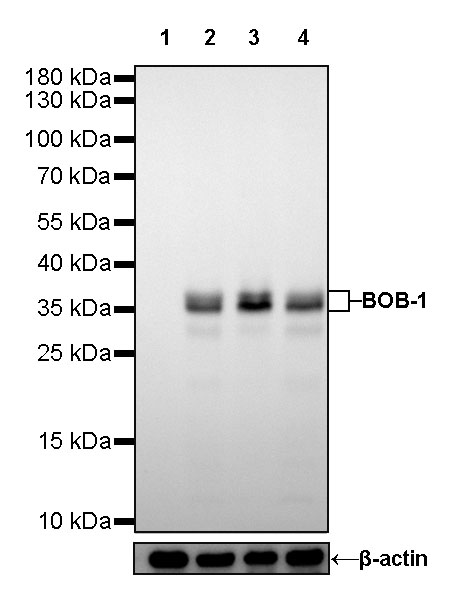
WB result of BOB-1 Rabbit mAb Primary antibody: BOB-1 Rabbit mAb at 1/5000 dilution Lane 1: HeLa whole cell lysate 20 µg Lane 2: Raji whole cell lysate 20 µg Lane 3: Ramos whole cell lysate 20 µg Lane 4: Daudi whole cell lysate 20 µg Negative control: HeLa whole cell lysate Secondary antibody: #JP20040 at 1/10000 dilution Predicted MW: 27 kDa Observed MW: 37 kDa Exposure time: 30s
IHC shows positive staining in paraffin-embedded human tonsil. Anti-BOB-1 antibody was used at 1/500 dilution, followed by a HRP Polymer for Mouse & Rabbit IgG (ready to use). Counterstained with hematoxylin. Heat mediated antigen retrieval with Tris/EDTA buffer pH9.0 was performed before commencing with IHC staining protocol.

IHC shows positive staining in paraffin-embedded human colon. Anti-BOB-1 antibody was used at 1/500 dilution, followed by a HRP Polymer for Mouse & Rabbit IgG (ready to use). Counterstained with hematoxylin. Heat mediated antigen retrieval with Tris/EDTA buffer pH9.0 was performed before commencing with IHC staining protocol.
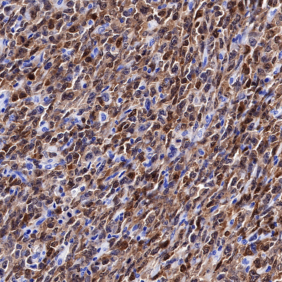
IHC shows positive staining in paraffin-embedded human diffuse large B-cell lymphoma. Anti-BOB-1 antibody was used at 1/500 dilution, followed by a HRP Polymer for Mouse & Rabbit IgG (ready to use). Counterstained with hematoxylin. Heat mediated antigen retrieval with Tris/EDTA buffer pH9.0 was performed before commencing with IHC staining protocol.
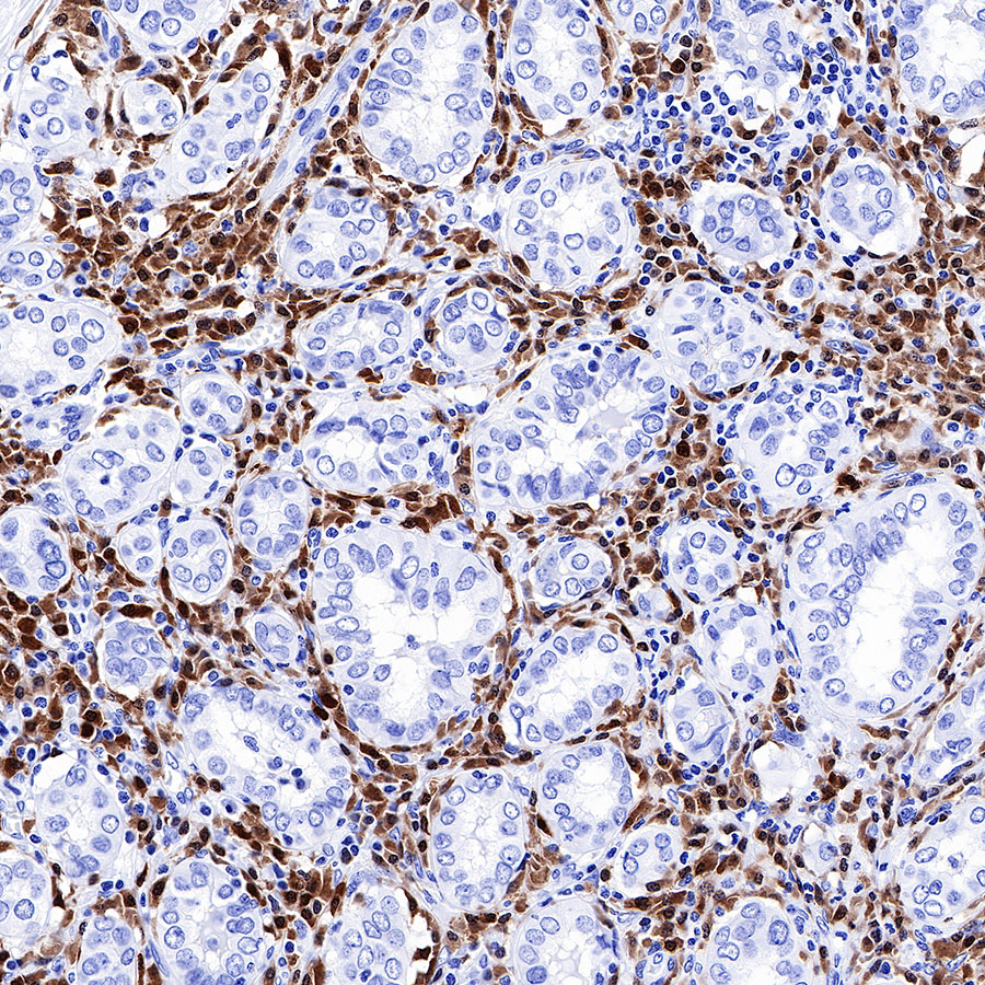
IHC shows positive staining in paraffin-embedded human thyroid cancer. Anti-BOB-1 antibody was used at 1/500 dilution, followed by a HRP Polymer for Mouse & Rabbit IgG (ready to use). Counterstained with hematoxylin. Heat mediated antigen retrieval with Tris/EDTA buffer pH9.0 was performed before commencing with IHC staining protocol.
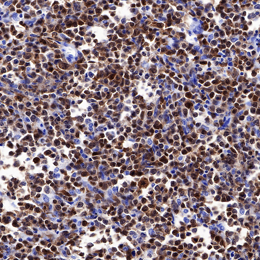
IHC shows positive staining in paraffin-embedded human diffuse large B-cell lymphoma. Anti-BOB-1 antibody was used at 1/500 dilution, followed by a HRP Polymer for Mouse & Rabbit IgG (ready to use). Counterstained with hematoxylin. Heat mediated antigen retrieval with Tris/EDTA buffer pH9.0 was performed before commencing with IHC staining protocol.
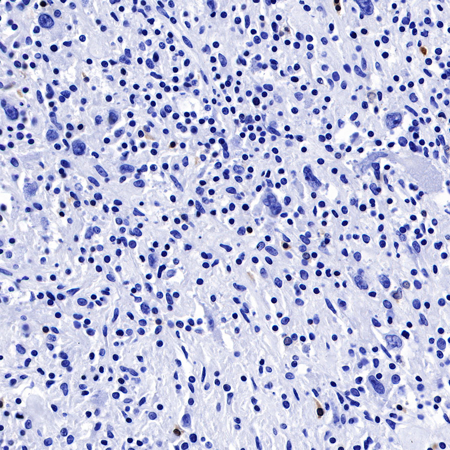
Negative control: IHC shows negative staining in paraffin-embedded human Hodgkin’s lymphoma. Anti-BOB-1 antibody was used at 1/500 dilution, followed by a HRP Polymer for Mouse & Rabbit IgG (ready to use). Counterstained with hematoxylin. Heat mediated antigen retrieval with Tris/EDTA buffer pH9.0 was performed before commencing with IHC staining protocol.
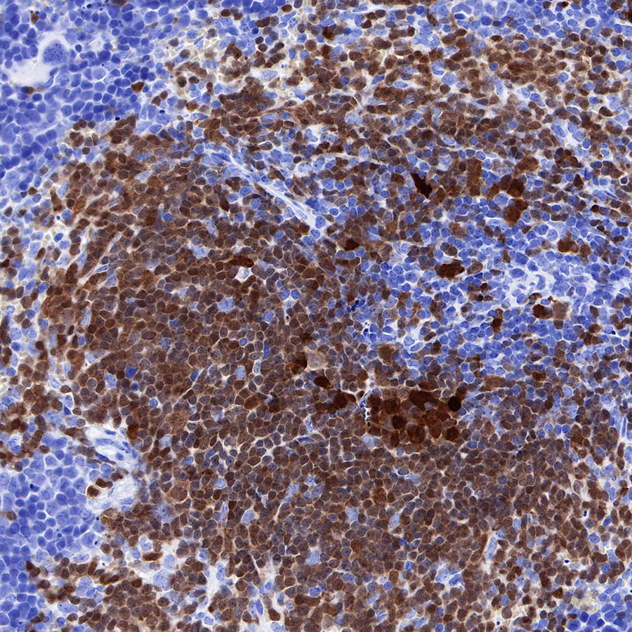
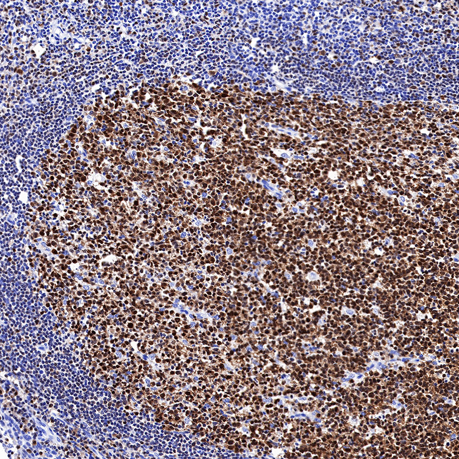
IHC shows positive staining in paraffin-embedded mouse spleen. Anti-BOB-1 antibody was used at 1/500 dilution, followed by a HRP Polymer for Mouse & Rabbit IgG (ready to use). Counterstained with hematoxylin. Heat mediated antigen retrieval with Tris/EDTA buffer pH9.0 was performed before commencing with IHC staining protocol.
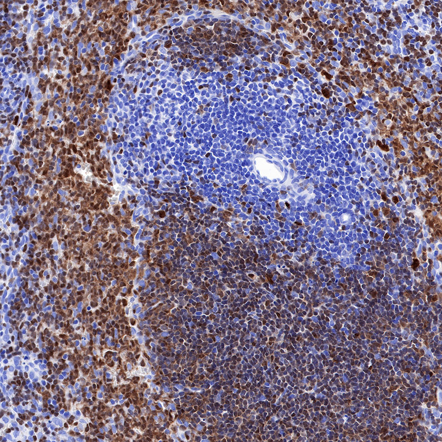
IHC shows positive staining in paraffin-embedded rat spleen. Anti-BOB-1 antibody was used at 1/500 dilution, followed by a HRP Polymer for Mouse & Rabbit IgG (ready to use). Counterstained with hematoxylin. Heat mediated antigen retrieval with Tris/EDTA buffer pH9.0 was performed before commencing with IHC staining protocol.