
Rabbit anti-CD3 epsilon Recombinant Monoclonal Antibody(241-49)别名宿主反应种属应用免疫原形式浓度纯化方法类型克隆号储存/保存方法存储溶液背景说明细胞定位UniProt
| 概述 | |
| 别名 |
T-cell surface antigen T3/Leu-4 epsilon chain; CD3e; T3E; CD3E
|
| 宿主 |
Rabbit
|
| 反应种属 |
Human, Mouse, Rat
|
| 应用 |
WB: 1:500, IP: 1:25, IHC-P: 1:2000-1:4000
|
| 免疫原 |
Synthetic peptide
|
| 性能 | |
| 形式 |
Liquid
|
| 浓度 |
0.25 mg/ml
|
| 纯化方法 |
Protein A affinity column
|
| 类型 |
Monoclonal Antibody
|
| 克隆号 |
241-49
|
| 储存/保存方法 |
Store at -20℃ for one year.
|
| 存储溶液 |
PBS, 40% Glycerol, 0.05% BSA, 0.03% Proclin 300
|
| 靶标 | |
| 背景说明 |
CD3 (cluster of differentiation 3) is a protein complex and T cell co-receptor that is involved in activating both the cytotoxic T cell (CD8+ naive T cells) and T helper cells (CD4+ naive T cells). It is composed of four distinct chains. In mammals, the complex contains a CD3γ chain, a CD3δ chain, and two CD3ε chains. These chains associate with the T-cell receptor (TCR) and the CD3-zeta (ζ-chain) to generate an activation signal in T lymphocytes. The TCR, CD3-zeta, and the other CD3 molecules together constitute the TCR complex. The CD3–T cell receptor (TCR) complex plays a central role in the T-cell-mediated immunoresponse as it is involved in the recognition of antigens and subsequent signal transduction and activation of immunocompetent T lymphocytes. Because CD3 is required for T cell activation, drugs (often monoclonal antibodies) that target it are being investigated as immunosuppressant therapies (e.g., otelixizumab, teplizumab) for type 1 diabetes and other autoimmune diseases.
|
| 细胞定位 |
Cell membrane
|
| UniProt |
P07766
|
实验结果图
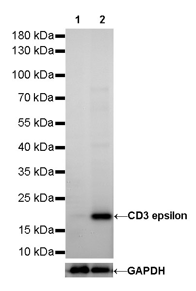
WB result of CD3 epsilon Rabbit mAb Primary antibody: CD3 epsilon Rabbit mAb at 1/500 dilution Lane 1: Raji whole cell lysate 20 µg Lane 2: Jurkat whole cell lysate 20 µg Negative control: Raji whole cell lysate Secondary antibody: #JP20040 at 1/10000 dilution Predicted MW: 20 kDa Observed MW: 20 kDa Exposure time: 13s
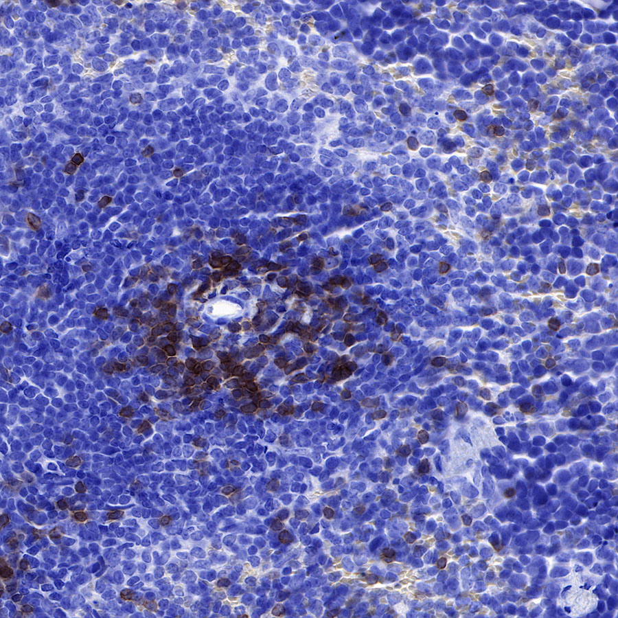
IHC shows positive staining in paraffin-embedded human spleen. Anti-CD3 epsilon antibody was used at 1/2000 dilution, Secondary antibody: #JP20040. Counterstained with hematoxylin. Heat mediated antigen retrieval with Tris/EDTA buffer pH9.0 was performed before commencing with IHC staining protocol.
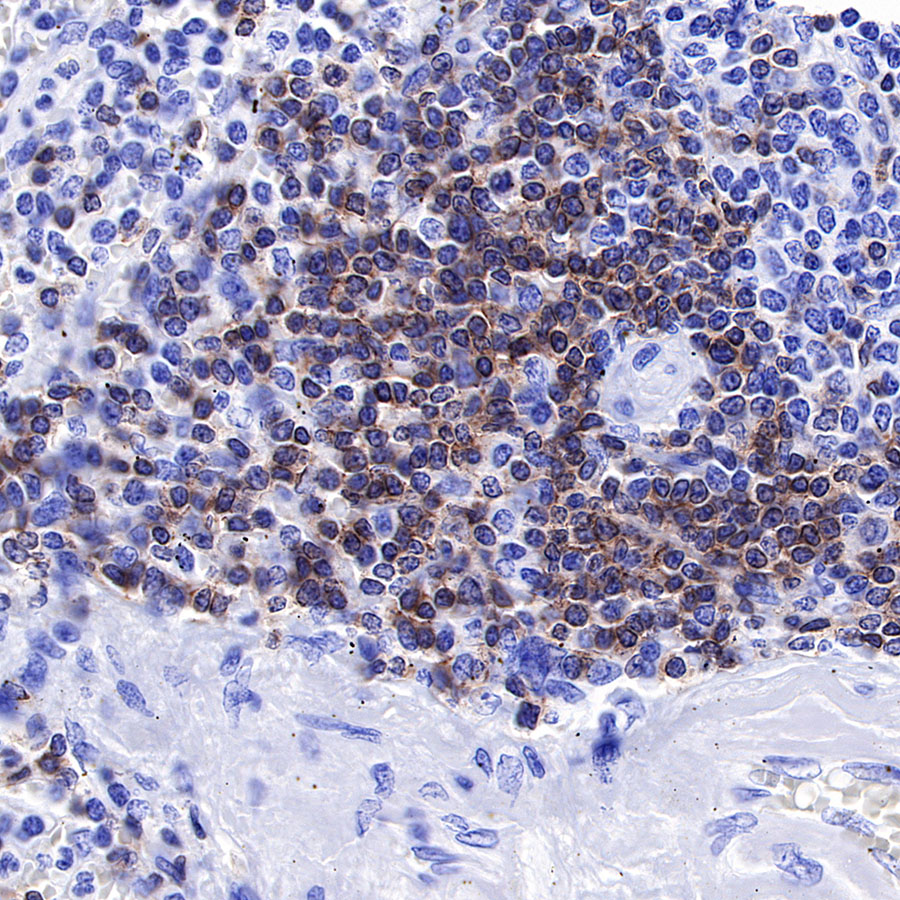
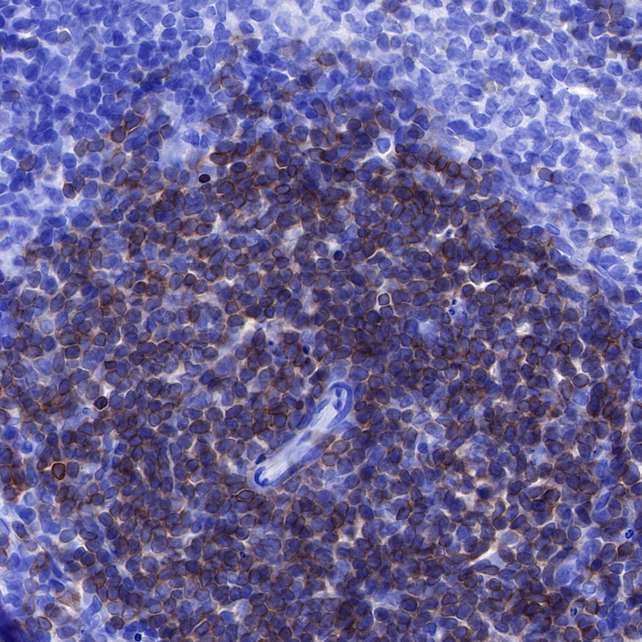
IHC shows positive staining in paraffin-embedded human tonsil. Anti-CD3 epsilon antibody was used at 1/2000 dilution, Secondary antibody: #JP20040. Counterstained with hematoxylin. Heat mediated antigen retrieval with Tris/EDTA buffer pH9.0 was performed before commencing with IHC staining protocol.
IHC shows positive staining in paraffin-embedded human tonsil. Anti-CD3 epsilon antibody was used at 1/2000 dilution, Secondary antibody: #JP20040. Counterstained with hematoxylin. Heat mediated antigen retrieval with Tris/EDTA buffer pH9.0 was performed before commencing with IHC staining protocol.
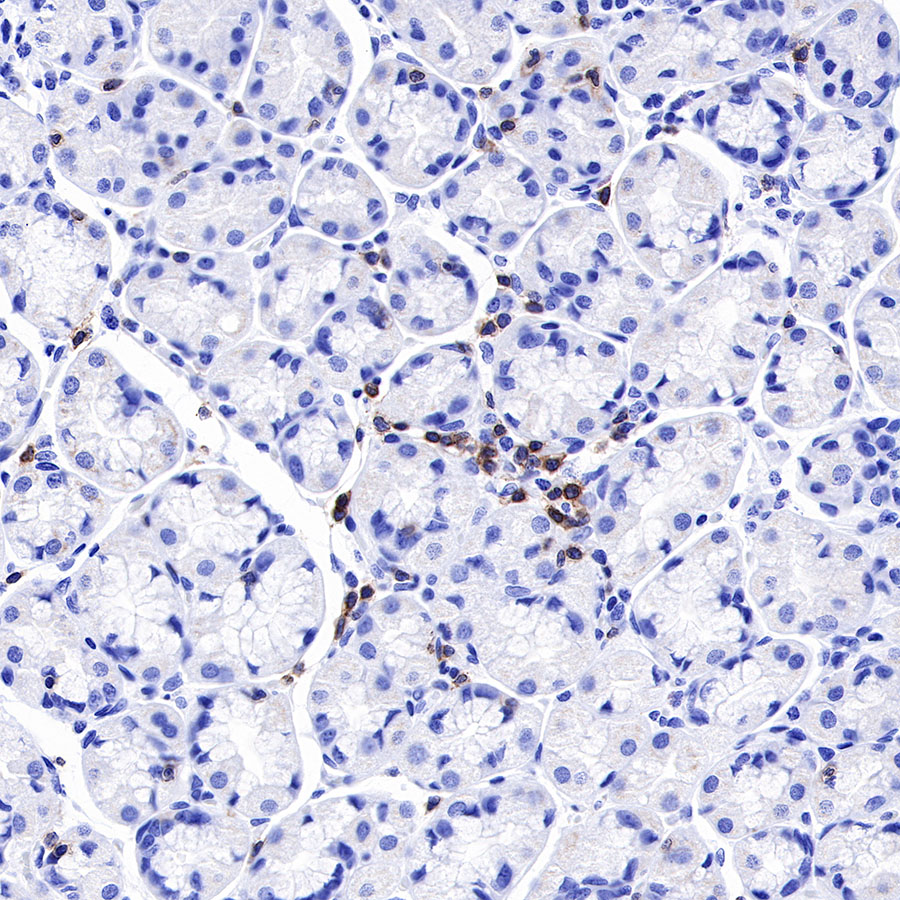
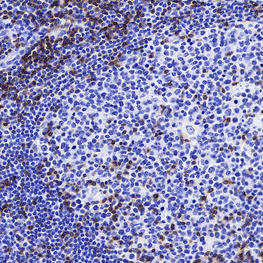
IHC shows positive staining in paraffin-embedded human stomach. Anti-CD3 epsilon antibody was used at 1/2000 dilution, Secondary antibody: #JP20040. Counterstained with hematoxylin. Heat mediated antigen retrieval with Tris/EDTA buffer pH9.0 was performed before commencing with IHC staining protocol.
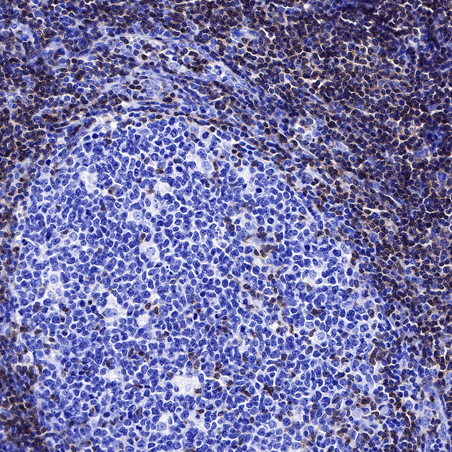
IHC shows positive staining in paraffin-embedded mouse spleen. Anti-CD3 epsilon antibody was used at 1/4000 dilution, Secondary antibody: #JP20040. Counterstained with hematoxylin. Heat mediated antigen retrieval with Tris/EDTA buffer pH9.0 was performed before commencing with IHC staining protocol.
IHC shows positive staining in paraffin-embedded rat spleen. Anti-CD3 epsilon antibody was used at 1/4000 dilution, Secondary antibody: #JP20040. Counterstained with hematoxylin. Heat mediated antigen retrieval with Tris/EDTA buffer pH9.0 was performed before commencing with IHC staining protocol.
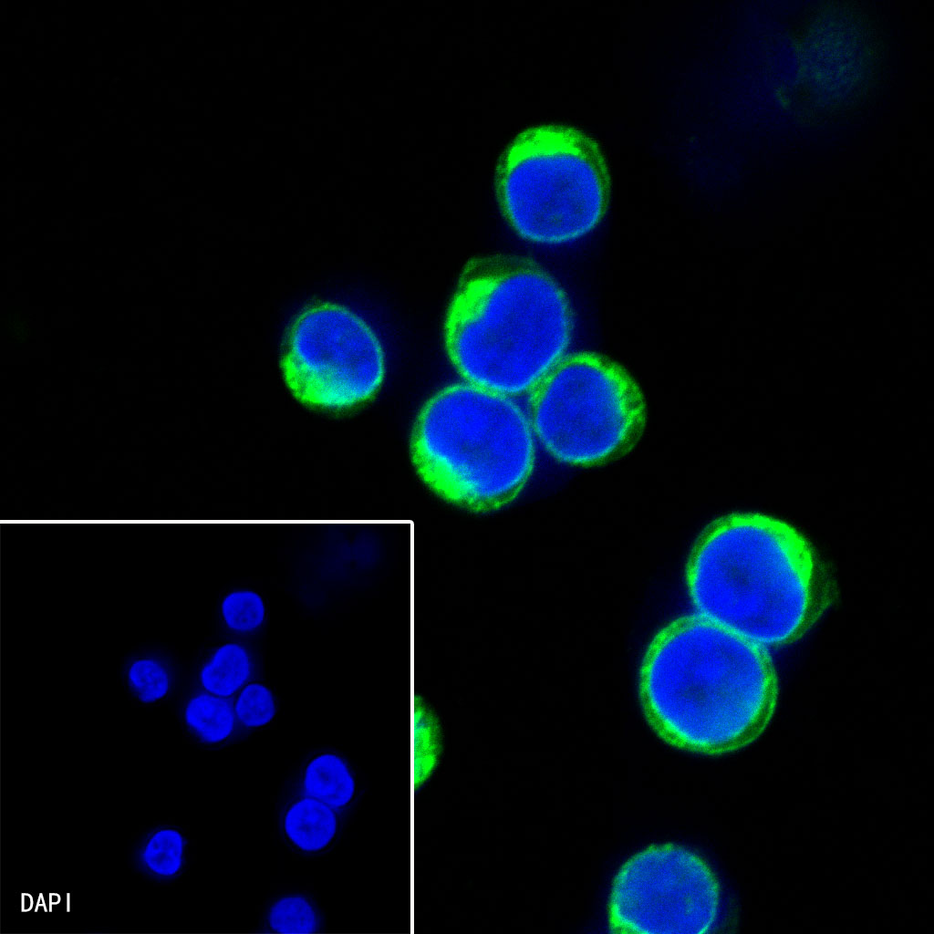
ICC shows positive staining in Jurkat cells. Anti-CD3 epsilon antibody was used at 1/500 dilution (Green) and incubated overnight at 4°C. Goat polyclonal Antibody to Rabbit IgG – H&L (Alexa Fluor® 488) was used as secondary antibody at 1/1000 dilution. The cells were fixed with 100% ice-cold methanol and permeabilized with 0.1% PBS-Triton X-100. Nuclei were counterstained with DAPI (Blue).
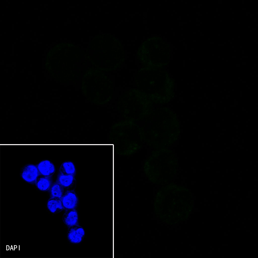
Negative control: ICC shows negative staining in Raji cells. Anti-CD3 epsilon antibody was used at 1/500 dilution (Green) and incubated overnight at 4°C. Goat polyclonal Antibody to Rabbit IgG – H&L (Alexa Fluor® 488) was used as secondary antibody at 1/1000 dilution. The cells were fixed with 100% ice-cold methanol and permeabilized with 0.1% PBS-Triton X-100. Nuclei were counterstained with DAPI (Blue).
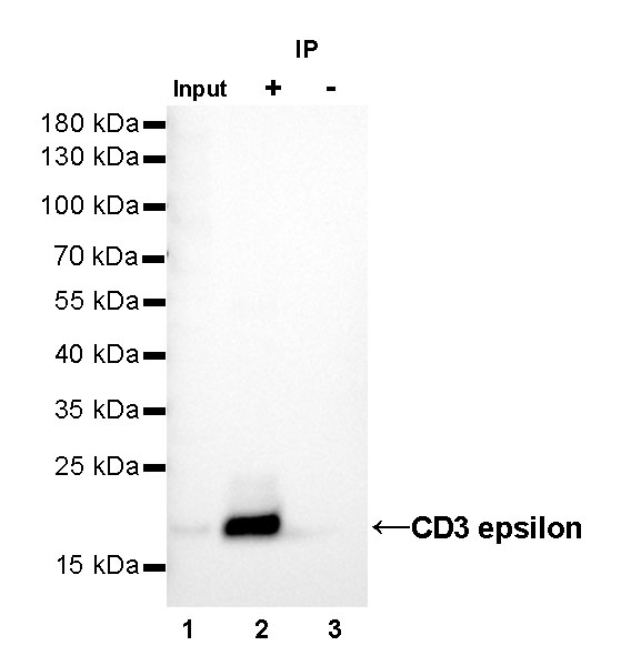
CD3 epsilon Rabbit mAb at 1/25 dilution (1µg) immunoprecipitating CD3 epsilon in 0.4mg Jurkat whole cell lysate. Western blot was performed on the immunoprecipitate using CD3 epsilon Rabbit mAb at 1/1000 dilution. Secondary antibody (HRP) for IP was used at 1/400 dilution. Lane 1 : Jurkat whole cell lysate 10µg(input) Lane 2 : CD3 epsilon Rabbit mAb IP in Jurkat whole cell lysate Lane 3 : Rabbit monoclonal IgG IP in Jurkat whole cell lysate Predicted MW: 20 kDa Observed MW: 20 kDa Exposure time: 10s