
Rabbit anti-IGFBP1 Recombinant Monoclonal Antibody(196-47)别名宿主反应种属应用免疫原形式浓度纯化方法类型克隆号储存/保存方法存储溶液背景说明细胞定位UniProt
| 概述 | |
| 别名 |
Insulin-like growth factor-binding protein 1, IBP-1; IGF-binding protein 1, Placental protein 12, PP12
|
| 宿主 |
Rabbit
|
| 反应种属 |
Human
|
| 应用 |
WB: 1:1000, ICC: 1:250, FC(Intra): 1:500
|
| 免疫原 |
Recombinant protein
|
| 性能 | |
| 形式 |
Liquid
|
| 浓度 |
0.5 mg/mL
|
| 纯化方法 |
Protein A affinity column
|
| 类型 |
Monoclonal Antibody
|
| 克隆号 |
196-47
|
| 储存/保存方法 |
Store at -20℃ for one year.
|
| 存储溶液 |
PBS, 40% Glycerol, 0.05% BSA, 0.03% Proclin 300
|
| 靶标 | |
| 背景说明 |
Insulin-like growth factor binding proteins (IGFBPs) are a family of proteins binding to Insulin-like growth factors (IGFs), generally including IGFBP1, IGFBP2, IGFBP3, IGFBP4, IGFBP5, and IGFBP6 [PMID: 30214426]. IGFBP1, a 40 to 50 kDa protein, which plays an indispensable role in normal growth and development, and in the pathophysiology of various tumors. IGFBP-1 has been shown to be associated with the risk of various tumors, and has a vital function in regulating tumor behaviors such as proliferation, migration, invasion and adhesion through different molecular mechanisms. The biological actions of IGFBP-1 in cancer are found to be related to its phosphorylation state, and the IGF-dependent and -independent mechanisms. [PMID: 1648311, PMID: 33841624].
|
| 细胞定位 |
Secreted
|
| UniProt |
P08833
|
实验结果图
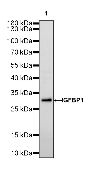
WB result of IGFBP1 Rabbit mAb Primary antibody: IGFBP1 Rabbit mAb at 1/1000 dilution Lane 1: HepG2 whole cell lysate 20 µg Secondary antibody: Goat Anti-Rabbit IgG, (H+L), HRP conjugated at 1/10000 dilution Predicted MW: 28 kDa Observed MW: 30 kDa Exposure time: 90s
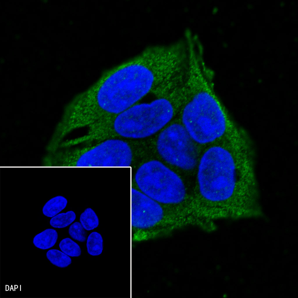
ICC shows positive staining in HepG2 cells. Anti-IGFBP1 antibody was used at 1/250 dilution (Green) and incubated overnight at 4°C. Goat polyclonal Antibody to Mouse IgG – H&L (Alexa Fluor® 488) was used as secondary antibody at 1/1000 dilution. The cells were fixed with 100% ice-cold methanol and permeabilized with 0.1% PBS-Triton X-100. Nuclei were counterstained with DAPI (Blue).
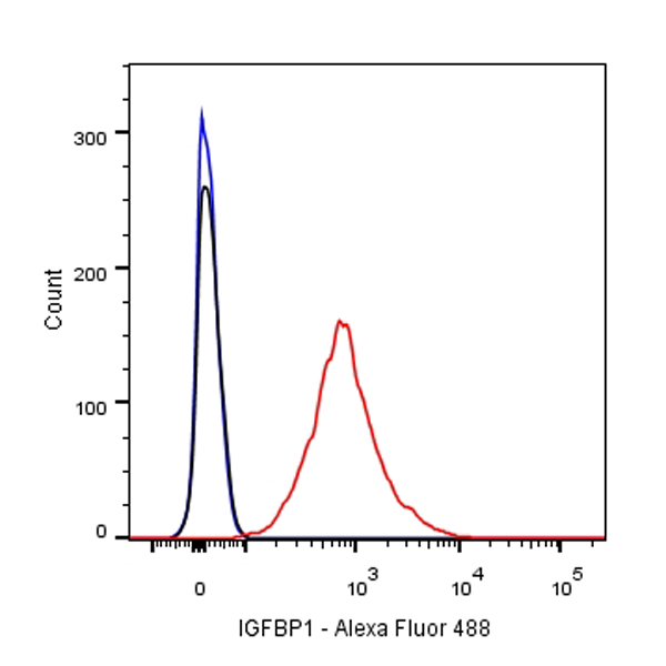
Flow cytometric analysis of HepG2 cells labelling IGFBP1 antibody at 1/500 (0.1 μg) dilution/ (red) compared with a Rabbit monoclonal IgG (Black) isotype control and an unlabelled control (cells without incubation with primary antibody and secondary antibody) (Blue). Goat Anti-Rabbit IgG Alexa Fluor® 488 was used as the secondary antibody.