
Rabbit anti-CD19 Recombinant Monoclonal Antibody(164-33)描述别名宿主特异性反应种属应用免疫原形式浓度纯化方法类型克隆号储存/保存方法存储溶液背景说明翻译后修饰细胞定位UniProt
| 概述 | |
| 描述 |
Lymphocytes proliferate and differentiate in response to various concentrations of different antigens. The ability of the B cell to respond in a specific, yet sensitive manner to the various antigens is achieved with the use of low-affinity antigen receptors. This gene encodes a cell surface molecule which assembles with the antigen receptor of B lymphocytes in order to decrease the threshold for antigen receptor-dependent stimulation.
|
| 别名 |
CD19抗体;B-lymphocyte antigen CD19; LE-CD19; B-lymphocyte surface antigen B4; Differentiation antigen CD19; T-cell surface antigen Leu-12
|
| 宿主 |
Rabbit
|
| 特异性 |
CD19 Antibody detects endogenous levels of total CD19.
|
| 反应种属 |
Human
|
| 应用 |
WB: 1:500, IHC-P: 1:1000, ICC: 1:250
|
| 免疫原 |
Synthetic peptide
|
| 性能 | |
| 形式 |
Liquid
|
| 浓度 |
0.25 mg/mL
|
| 纯化方法 |
Protein A affinity column
|
| 类型 |
Monoclonal Antibody
|
| 克隆号 |
164-33
|
| 储存/保存方法 |
Store at -20℃ for one year.
|
| 存储溶液 |
PBS, 40% Glycerol, 0.05% BSA, 0.03% Proclin 300
|
| 靶标 | |
| 背景说明 |
The CD19 antigen plays an important role in clinical oncology. In normal cells, it is the most ubiquitously expressed protein in the B lymphocyte lineage. CD19 expression is induced at the point of B lineage commitment during the differentiation of the hematopoietic stem cell, and its expression continues through preB and mature B cell differentiation until it is finally down-regulated during terminal differentiation into plasma cells. CD19 expression is maintained in B-lineage cells that have undergone neoplastic transformation, and therefore CD19 is useful in diagnosis of leukemias and lymphomas using monoclonal antibodies (mAbs) and flow cytometry [PMID: 8528044].
|
| 翻译后修饰 |
Phosphorylated on serine and threonine upon DNA damage, probably by ATM or ATR. Phosphorylated on tyrosine following B-cell activation. Phosphorylated on tyrosine residues by LYN.
|
| 细胞定位 |
Cell Membrane
|
| UniProt |
P15391
|
实验结果图
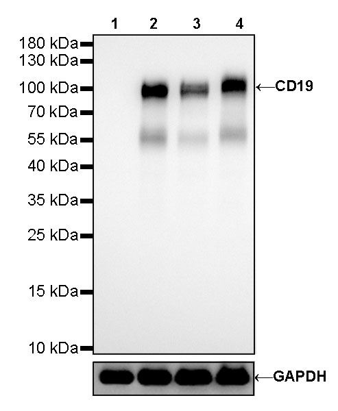
WB result of CD19 Rabbit mAb Primary antibody: CD19 Rabbit mAb at 1/500 dilution Lane 1: Jurkat whole cell lysate 20ug Lane 2: Raji whole cell lysate 20ug Lane 3: Ramos whole cell lysate 20ug Lane 4: Daudi whole cell lysate 20ug Negative control: Jurkat whole cell lysate Secondary antibody: #JP20040 at 1/10000 dilution Predicted MW: 95 kDa Observed MW: 95 kDa Exposure time: 30s

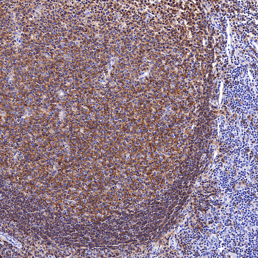
IHC shows positive staining in paraffin-embedded human spleen. Anti-CD19 antibody was used at 1/1000 dilution, followed by a HRP Polymer for Mouse & Rabbit IgG (ready to use). Counterstained with hematoxylin. Heat mediated antigen retrieval with Tris/EDTA buffer pH9.0 was performed before commencing with IHC staining protocol.
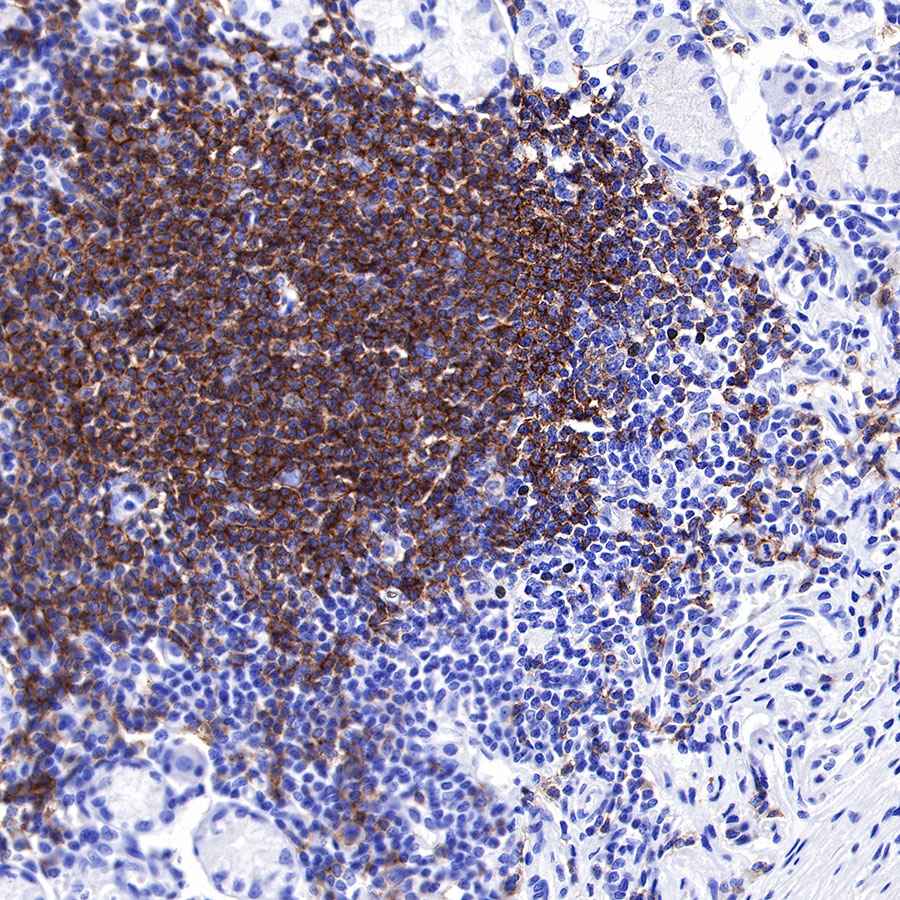
IHC shows positive staining in paraffin-embedded human tonsil. Anti-CD19 antibody was used at 1/1000 dilution, followed by a HRP Polymer for Mouse & Rabbit IgG (ready to use). Counterstained with hematoxylin. Heat mediated antigen retrieval with Tris/EDTA buffer pH9.0 was performed before commencing with IHC staining protocol.
IHC shows positive staining in paraffin-embedded human stomach. Anti-CD19 antibody was used at 1/1000 dilution, followed by a HRP Polymer for Mouse & Rabbit IgG (ready to use). Counterstained with hematoxylin. Heat mediated antigen retrieval with Tris/EDTA buffer pH9.0 was performed before commencing with IHC staining protocol.
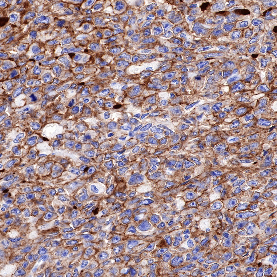
IHC shows positive staining in paraffin-embedded human diffuse large B-cell lymphoma. Anti-CD19 antibody was used at 1/1000 dilution, followed by a HRP Polymer for Mouse & Rabbit IgG (ready to use). Counterstained with hematoxylin. Heat mediated antigen retrieval with Tris/EDTA buffer pH9.0 was performed before commencing with IHC staining protocol.
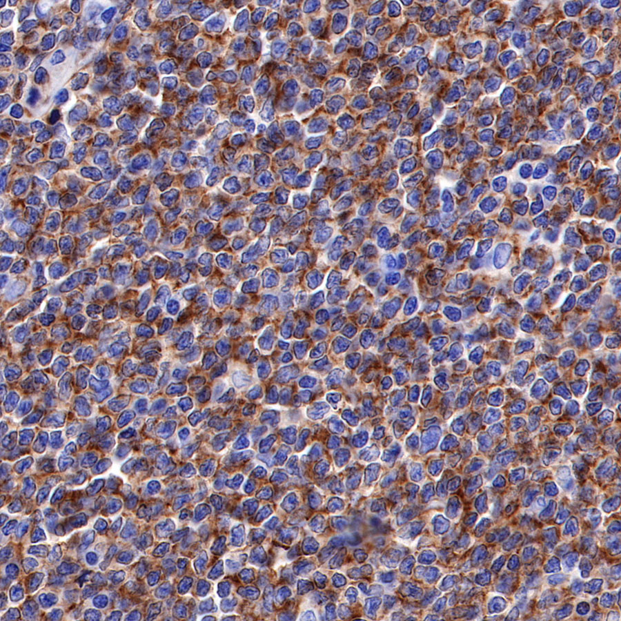
IHC shows positive staining in paraffin-embedded human mantle cell lymphoma. Anti-CD19 antibody was used at 1/1000 dilution, followed by a HRP Polymer for Mouse & Rabbit IgG (ready to use). Counterstained with hematoxylin. Heat mediated antigen retrieval with Tris/EDTA buffer pH9.0 was performed before commencing with IHC staining protocol.
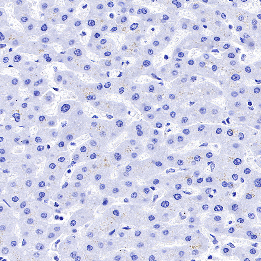
Negative control: IHC shows negative staining in paraffin-embedded human liver. Anti-CD19 antibody was used at 1/1000 dilution, followed by a HRP Polymer for Mouse & Rabbit IgG (ready to use). Counterstained with hematoxylin. Heat mediated antigen retrieval with Tris/EDTA buffer pH9.0 was performed before commencing with IHC staining protocol.
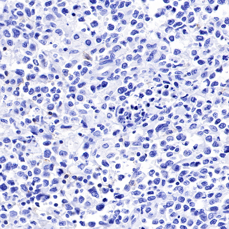
Negative control: IHC shows negative staining in paraffin-embedded human NK/T-cell lymphoma. Anti-CD19 antibody was used at 1/1000 dilution, followed by a HRP Polymer for Mouse & Rabbit IgG (ready to use). Counterstained with hematoxylin. Heat mediated antigen retrieval with Tris/EDTA buffer pH9.0 was performed before commencing with IHC staining protocol.
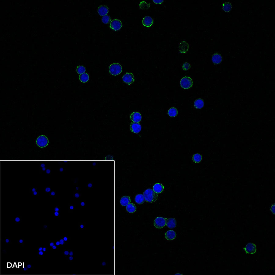
ICC shows positive staining in Ramos cells. Anti-CD19 antibody was used at 1/250 dilution and incubated overnight at 4°C. Goat polyclonal Antibody to Rabbit IgG – H&L (Alexa Fluor® 488) was used as secondary antibody at 1/1000 dilution. The cells were fixed with 100% Methanol and permeabilized with 0.1% PBS-Triton X-100. Nuclei were counterstained with DAPI.
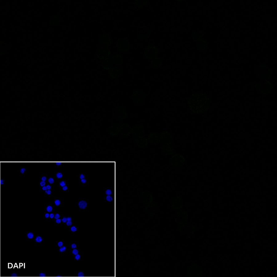
Negative control: ICC shows negative staining in Jurkat cells. Anti-CD19 antibody was used at 1/250 dilution and incubated overnight at 4°C. Goat polyclonal Antibody to Rabbit IgG – H&L (Alexa Fluor® 488) was used as secondary antibody at 1/1000 dilution. The cells were fixed with 100% Methanol and permeabilized with 0.1% PBS-Triton X-100. Nuclei were counterstained with DAPI.