
Rabbit anti-CD19 Recombinant Monoclonal Antibody(164-33)别名宿主反应种属应用免疫原形式浓度纯化方法类型克隆号储存/保存方法存储溶液背景说明细胞定位UniProt
| 概述 | |
| 别名 |
B-lymphocyte antigen CD19; LE-CD19; B-lymphocyte surface antigen B4; Differentiation antigen CD19; T-cell surface antigen Leu-12
|
| 宿主 |
Rabbit
|
| 反应种属 |
Human
|
| 应用 |
WB: 1:500, IHC-P: 1:1000, ICC: 1:250
|
| 免疫原 |
Synthetic peptide
|
| 性能 | |
| 形式 |
Liquid
|
| 浓度 |
0.25 mg/mL
|
| 纯化方法 |
Protein A affinity column
|
| 类型 |
Monoclonal Antibody
|
| 克隆号 |
164-33
|
| 储存/保存方法 |
Store at -20℃ for one year.
|
| 存储溶液 |
PBS, 40% Glycerol, 0.05% BSA, 0.03% Proclin 300
|
| 靶标 | |
| 背景说明 |
The CD19 antigen plays an important role in clinical oncology. In normal cells, it is the most ubiquitously expressed protein in the B lymphocyte lineage. CD19 expression is induced at the point of B lineage commitment during the differentiation of the hematopoietic stem cell, and its expression continues through preB and mature B cell differentiation until it is finally down-regulated during terminal differentiation into plasma cells. CD19 expression is maintained in B-lineage cells that have undergone neoplastic transformation, and therefore CD19 is useful in diagnosis of leukemias and lymphomas using monoclonal antibodies (mAbs) and flow cytometry [PMID: 8528044].
|
| 细胞定位 |
Cell Membrane
|
| UniProt |
P15391
|
实验结果图
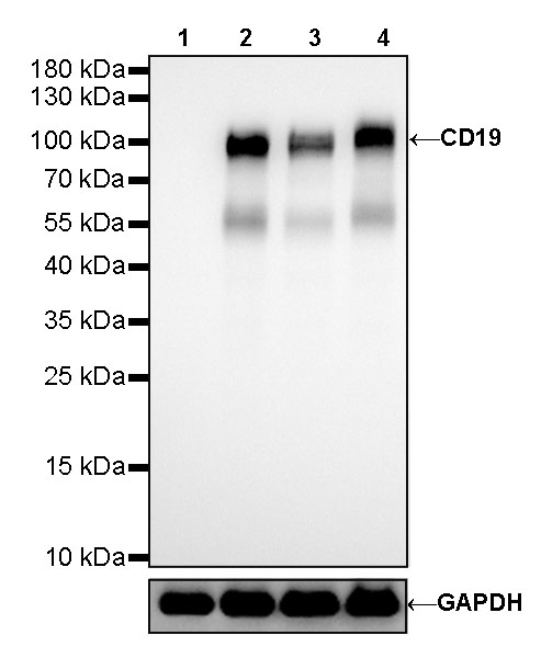
WB result of CD19 Rabbit mAb Primary antibody: CD19 Rabbit mAb at 1/500 dilution Lane 1: Jurkat whole cell lysate 20ug Lane 2: Raji whole cell lysate 20ug Lane 3: Ramos whole cell lysate 20ug Lane 4: Daudi whole cell lysate 20ug Negative control: Jurkat whole cell lysate Secondary antibody: #JP20040 at 1/10000 dilution Predicted MW: 95 kDa Observed MW: 95 kDa Exposure time: 30s

IHC shows positive staining in paraffin-embedded human spleen. Anti-CD19 antibody was used at 1/1000 dilution, followed by a HRP Polymer for Mouse & Rabbit IgG (ready to use). Counterstained with hematoxylin. Heat mediated antigen retrieval with Tris/EDTA buffer pH9.0 was performed before commencing with IHC staining protocol.
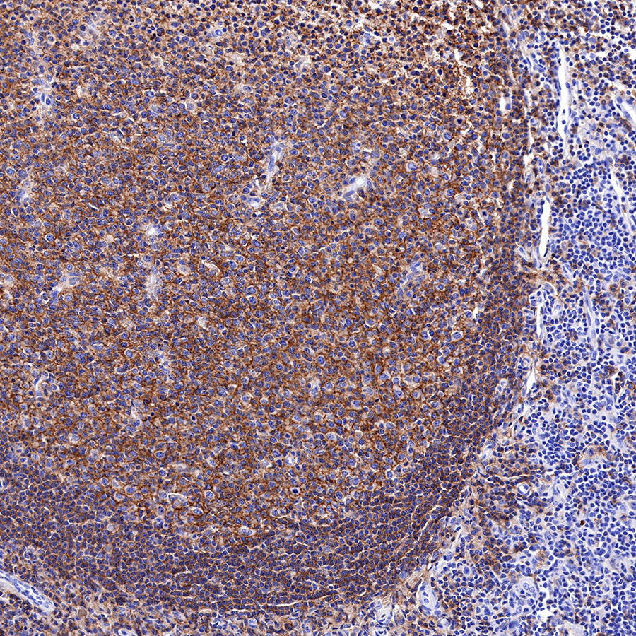
IHC shows positive staining in paraffin-embedded human tonsil. Anti-CD19 antibody was used at 1/1000 dilution, followed by a HRP Polymer for Mouse & Rabbit IgG (ready to use). Counterstained with hematoxylin. Heat mediated antigen retrieval with Tris/EDTA buffer pH9.0 was performed before commencing with IHC staining protocol.
IHC shows positive staining in paraffin-embedded human stomach. Anti-CD19 antibody was used at 1/1000 dilution, followed by a HRP Polymer for Mouse & Rabbit IgG (ready to use). Counterstained with hematoxylin. Heat mediated antigen retrieval with Tris/EDTA buffer pH9.0 was performed before commencing with IHC staining protocol.
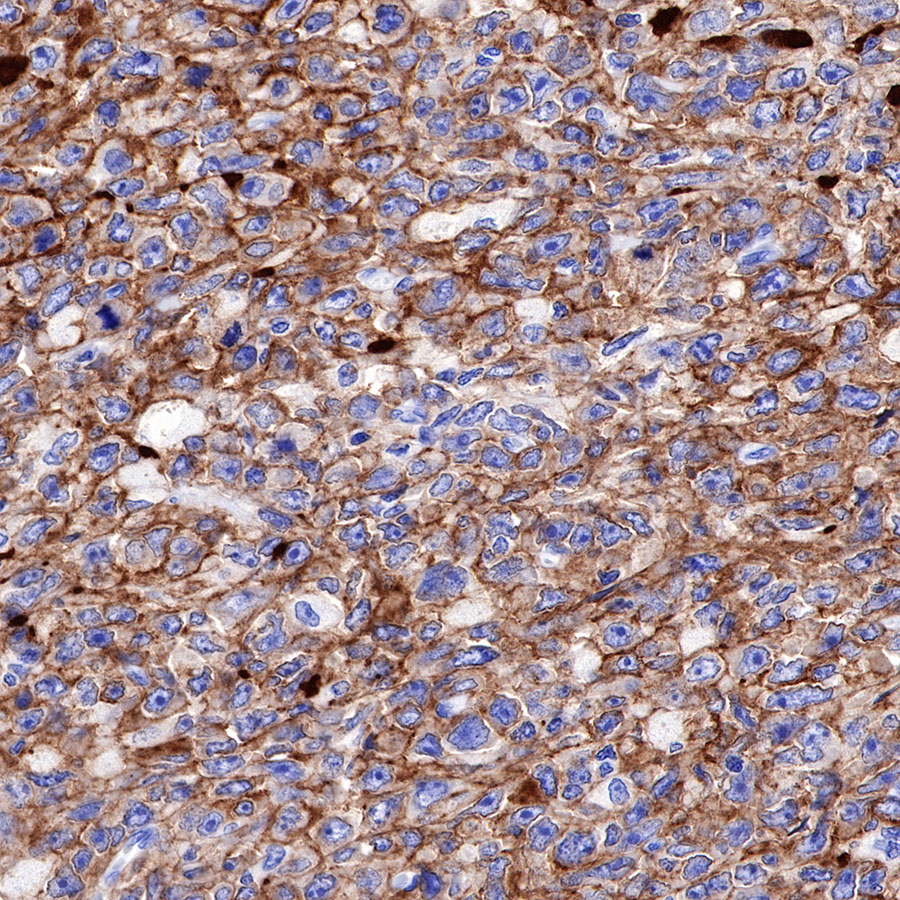
IHC shows positive staining in paraffin-embedded human diffuse large B-cell lymphoma. Anti-CD19 antibody was used at 1/1000 dilution, followed by a HRP Polymer for Mouse & Rabbit IgG (ready to use). Counterstained with hematoxylin. Heat mediated antigen retrieval with Tris/EDTA buffer pH9.0 was performed before commencing with IHC staining protocol.
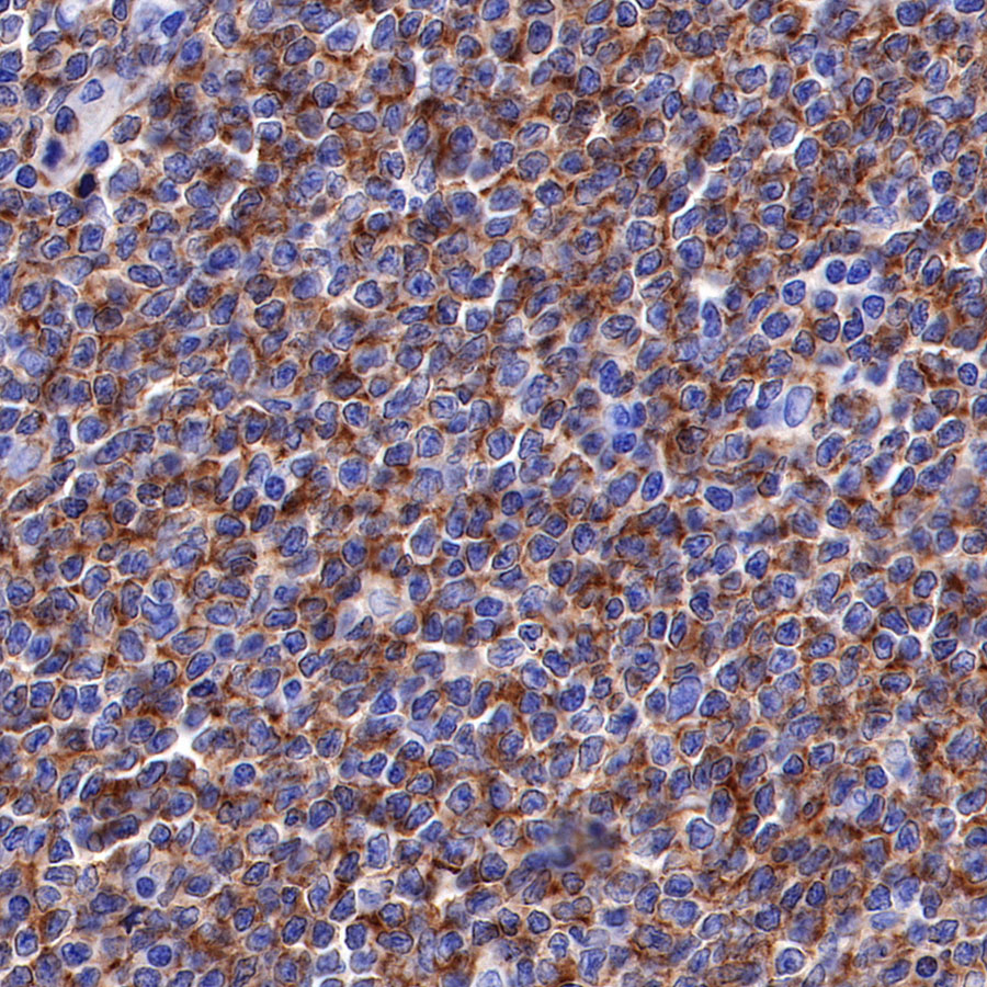
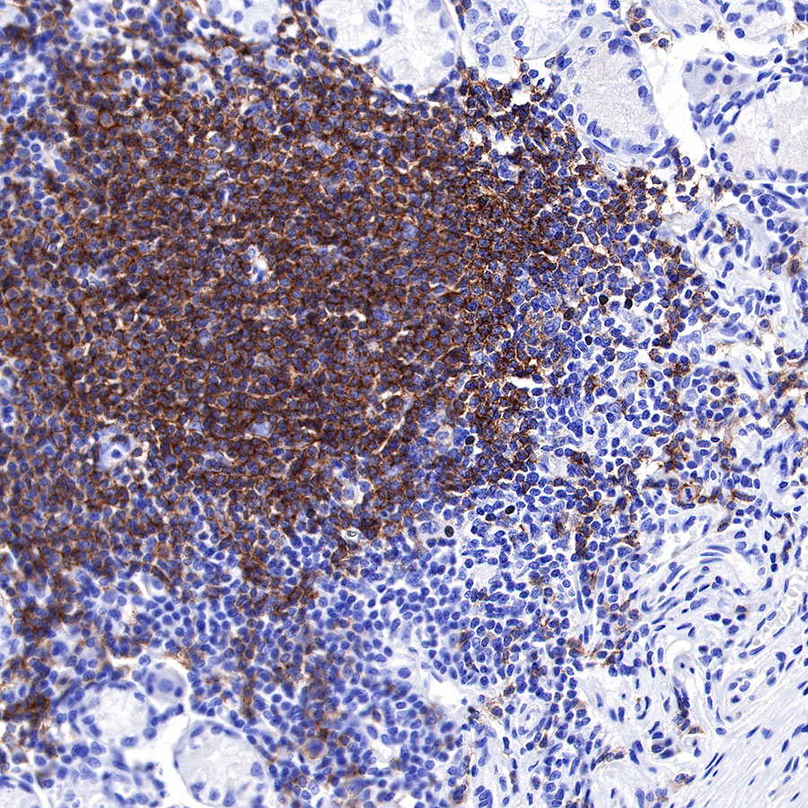
IHC shows positive staining in paraffin-embedded human mantle cell lymphoma. Anti-CD19 antibody was used at 1/1000 dilution, followed by a HRP Polymer for Mouse & Rabbit IgG (ready to use). Counterstained with hematoxylin. Heat mediated antigen retrieval with Tris/EDTA buffer pH9.0 was performed before commencing with IHC staining protocol.
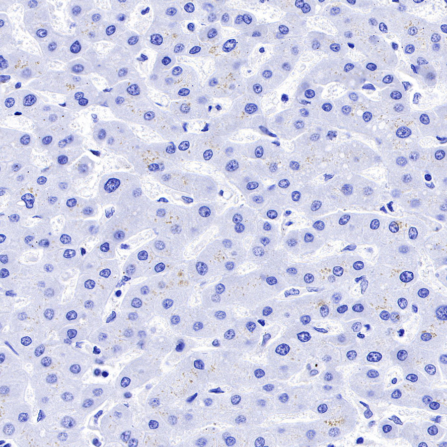
Negative control: IHC shows negative staining in paraffin-embedded human liver. Anti-CD19 antibody was used at 1/1000 dilution, followed by a HRP Polymer for Mouse & Rabbit IgG (ready to use). Counterstained with hematoxylin. Heat mediated antigen retrieval with Tris/EDTA buffer pH9.0 was performed before commencing with IHC staining protocol.
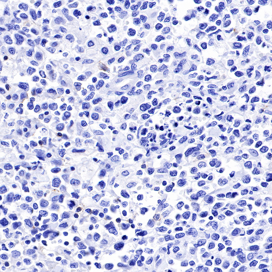
Negative control: IHC shows negative staining in paraffin-embedded human NK/T-cell lymphoma. Anti-CD19 antibody was used at 1/1000 dilution, followed by a HRP Polymer for Mouse & Rabbit IgG (ready to use). Counterstained with hematoxylin. Heat mediated antigen retrieval with Tris/EDTA buffer pH9.0 was performed before commencing with IHC staining protocol.
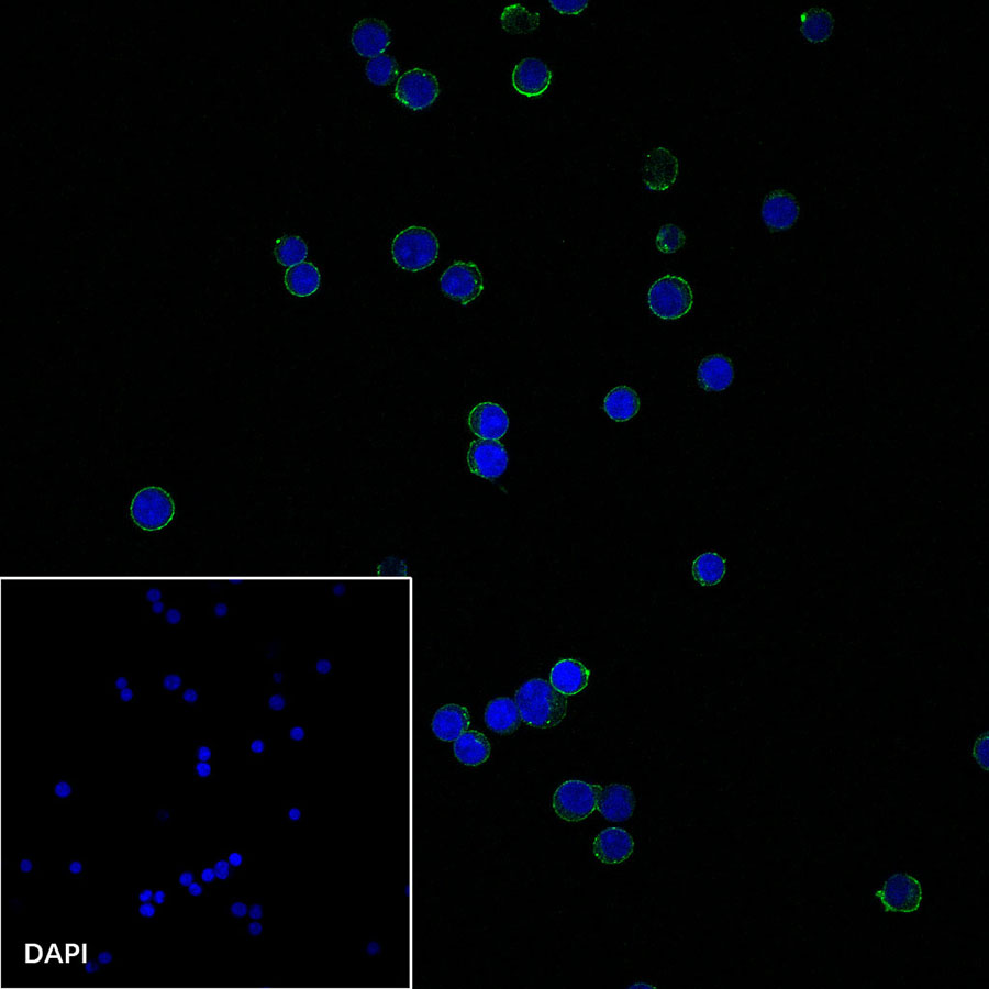
ICC shows positive staining in Ramos cells. Anti-CD19 antibody was used at 1/250 dilution and incubated overnight at 4°C. Goat polyclonal Antibody to Rabbit IgG – H&L (Alexa Fluor® 488) was used as secondary antibody at 1/1000 dilution. The cells were fixed with 100% Methanol and permeabilized with 0.1% PBS-Triton X-100. Nuclei were counterstained with DAPI.
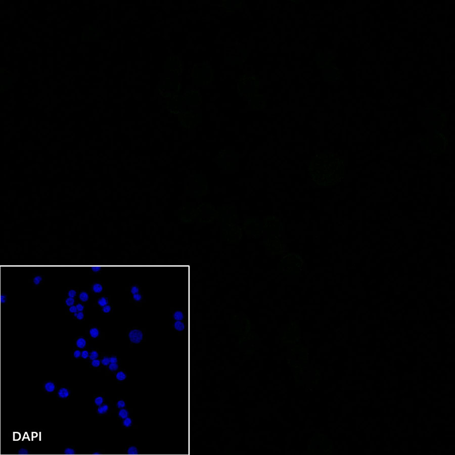
Negative control: ICC shows negative staining in Jurkat cells. Anti-CD19 antibody was used at 1/250 dilution and incubated overnight at 4°C. Goat polyclonal Antibody to Rabbit IgG – H&L (Alexa Fluor® 488) was used as secondary antibody at 1/1000 dilution. The cells were fixed with 100% Methanol and permeabilized with 0.1% PBS-Triton X-100. Nuclei were counterstained with DAPI.