
Mouse anti-AURKA Monoclonal Antibody(1364CT291.108.155)描述别名宿主特异性反应种属应用分子量类型克隆号同种型储存/保存方法研究领域背景说明细胞定位UniProt参考文献
| 概述 | |
| 描述 |
Purified Mouse Monoclonal Antibody (Mab)
|
| 别名 |
AURKA抗体;Aurora kinase A; Aurora 2; Aurora/IPL1-related kinase 1; ARK-1; Aurora-related kinase 1; hARK1; Breast tumor-amplified kinase; Serine/threonine-protein kinase 15; Serine/threonine-protein kinase 6; Serine/threonine-protein kinase aurora-A; AURKA
|
| 宿主 |
Mouse
|
| 特异性 |
This AURKA antibody is generated from a mouse immunized with a KLH conjugated synthetic peptide between amino acids from the human region of human AURKA.
|
| 反应种属 |
Human
|
| 应用 |
IHC-P~~1:25
WB~~1:1000 |
| 分子量 |
Predicted molecular weight: 46kD
Disclaimer note: The observed molecular weight of the protein may vary from the listed predicted molecular weight due to post translational modifications, post translation cleavages, relative charges, and other experimental factors. |
| 性能 | |
| 类型 |
Monoclonal Antibody
|
| 克隆号 |
1364CT291.108.155
|
| 同种型 |
IgG1,k
|
| 储存/保存方法 |
Maintain refrigerated at 2-8°C for up to 2 weeks. For long term storage store at -20°C in small aliquots to prevent freeze-thaw cycles.
|
| 研究领域 |
Cancer;Cell Biology;Signal Transduction
|
| 靶标 | |
| 背景说明 |
Mitotic serine/threonine kinases that contributes to the regulation of cell cycle progression. Associates with the centrosome and the spindle microtubules during mitosis and plays a critical role in various mitotic events including the establishment of mitotic spindle, centrosome duplication, centrosome separation as well as maturation, chromosomal alignment, spindle assembly checkpoint, and cytokinesis. Required for initial activation of CDK1 at centrosomes. Phosphorylates numerous target proteins, including ARHGEF2, BORA, BRCA1, CDC25B, DLGP5, HDAC6, KIF2A, LATS2, NDEL1, PARD3, PPP1R2, PLK1, RASSF1, TACC3, p53/TP53 and TPX2. Regulates KIF2A tubulin depolymerase activity. Required for normal axon formation. Plays a role in microtubule remodeling during neurite extension. Important for microtubule formation and/or stabilization. Also acts as a key regulatory component of the p53/TP53 pathway, and particularly the checkpoint-response pathways critical for oncogenic transformation of cells, by phosphorylating and stabilizing p53/TP53. Phosphorylates its own inhibitors, the protein phosphatase type 1 (PP1) isoforms, to inhibit their activity. Necessary for proper cilia disassembly prior to mitosis.
|
| 细胞定位 |
Cytoplasm, cytoskeleton, microtubule organizing center, centrosome. Cytoplasm, cytoskeleton, spindle pole. Note=Detected at the neurite hillock in developing neurons (By similarity). Localizes at the centrosome in mitotic cells from early prophase until telophase, but also localizes to the spindle pole MTs from prophase to anaphase. Colocalized with SIRT2 at centrosome. Moves to the midbody during both telophase and cytokinesis. Associates with both the pericentriolar material (PCM) and centrioles.
|
| UniProt |
O14965
|
| 参考文献 | |
| 参考文献 |
Kimura M.,et al.J. Biol. Chem. 272:13766-13771(1997).
Shindo M.,et al.Biochem. Biophys. Res. Commun. 244:285-292(1998). Zhou H.,et al.Nat. Genet. 20:189-193(1998). Wang L.,et al.Submitted (OCT-1999) to the EMBL/GenBank/DDBJ databases. Deloukas P.,et al.Nature 414:865-871(2001). |
实验结果图
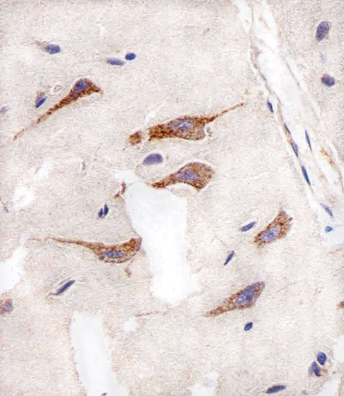
Immunohistochemical analysis of paraffin-embedded H. brain section using AURKA Antibody(Cat#JP100457). JP100457 was diluted at 1:25 dilution. A peroxidase-conjugated goat anti-mouse IgG at 1:400 dilution was used as the secondary antibody, followed by DAB staining.
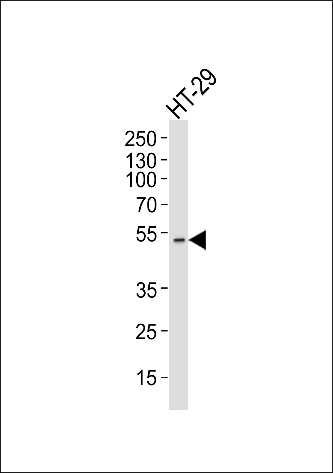
Western blot analysis of lysate from HT-29 cell line, using AURKA Antibody(Cat. #JP100457). JP100457 was diluted at 1:1000. A goat anti-mouse IgG H&L(HRP) at 1:3000 dilution was used as the secondary antibody. Lysate at 35μg.