
Rabbit anti-CD9 Recombinant Monoclonal Antibody(143-53)宿主反应种属应用分子量免疫原形式浓度纯化方法类型克隆号储存/保存方法存储溶液背景说明细胞定位UniProt
| 概述 | |
| 宿主 |
Rabbit
|
| 反应种属 |
Human
|
| 应用 |
WB: 1:1000, IP: 1:25, IHC-P: 1:500-1:2000, FC(Extra): 1:500
|
| 分子量 |
25 kDa
|
| 免疫原 |
Synthetic peptide
|
| 性能 | |
| 形式 |
liquid
|
| 浓度 |
0.5 mg/ml
|
| 纯化方法 |
Protein A affinity column
|
| 类型 |
Monoclonal antibody
|
| 克隆号 |
143-53
|
| 储存/保存方法 |
Store at -20℃ for one year.
|
| 存储溶液 |
PBS, 40% Glycerol, 0.05% BSA, 0.03% Proclin 300
|
| 靶标 | |
| 背景说明 |
D9 belongs to the cell surface glycoprotein cross -membrane four -protein family, with four cross -membrane domains, a shorter outer domain (ECL1), and a longer extracellular domain (ECL2). It is expressed in a variety of hematopoietic cells and epithelial cells. For example, Pre B cells, B cell subset, activated T cells, basophils, eosinophils, macrophages, megakaryocytes, plasma cells, plasma cell precursors in germinal centers, and platelets. Broadcasting a series of cell processes such as cell adhesion, movement, membrane tissue and signal transfusion. The lowering of CD9 expression is related to the adverse prognosis and progress of various cancers. It is a favorable marker of gallbladder cancer, gastic GIST, malignant cortex and oral squamous cell carcinoma.
|
| 细胞定位 |
Cell membrane
|
| UniProt |
P21926
|
实验结果图
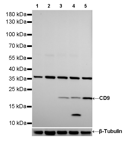
WB result of CD9 Rabbit mAb Primary antibody: CD9 Rabbit mAb at 1/1000 dilution Lane 1: Raji whole cell lysate 20 µg Lane 2: K562 whole cell lysate 20 µg Lane 3: Hela whole cell lysate 20 µg Lane 4: HCT 116 whole cell lysate 20 µg Lane 5: MCF7 whole cell lysate 20 µg Negative control: Raji whole cell lysate; K562 whole cell lysate Secondary antibody: #JP20040 at 1/10000 dilution Predicted MW: 22 kDa Observed MW: 22 kDa Exposure time: 180s

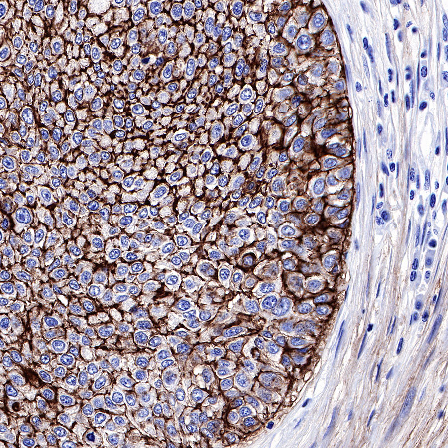
IHC shows positive staining in paraffin-embedded human tonsil.Anti-CD9 antibody was used at 1/500 dilution, Secondary antibody: #JP20040. Counterstained with hematoxylin. Heat mediated antigen retrieval with Tris/EDTA buffer pH9.0 was performed before commencing with IHC staining protocol.

IHC shows positive staining in paraffin-embedded human spleen.Anti-CD9 antibody was used at 1/500 dilution, Secondary antibody: #JP20040. Counterstained with hematoxylin. Heat mediated antigen retrieval with Tris/EDTA buffer pH9.0 was performed before commencing with IHC staining protocol.

IHC shows positive staining in paraffin-embedded human prostate.Anti-CD9 antibody was used at 1/500 dilution, Secondary antibody: #JP20040. Counterstained with hematoxylin. Heat mediated antigen retrieval with Tris/EDTA buffer pH9.0 was performed before commencing with IHC staining protocol.

IHC shows positive staining in paraffin-embedded human human ovarian cancer.Anti-CD9 antibody was used at 1/500 dilution, Secondary antibody: #JP20040. Counterstained with hematoxylin. Heat mediated antigen retrieval with Tris/EDTA buffer pH9.0 was performed before commencing with IHC staining protocol.
IHC shows positive staining in paraffin-embedded human human lung squamous cancer.Anti-CD9 antibody was used at 1/2000 dilution, Secondary antibody: #JP20040. Counterstained with hematoxylin. Heat mediated antigen retrieval with Tris/EDTA buffer pH9.0 was performed before commencing with IHC staining protocol.

IHC shows positive staining in paraffin-embedded human breast cancer.Anti-CD9 antibody was used at 1/2000 dilution, Secondary antibody: #JP20040. Counterstained with hematoxylin. Heat mediated antigen retrieval with Tris/EDTA buffer pH9.0 was performed before commencing with IHC staining protocol.

IHC shows positive staining in paraffin-embedded human bladder cancer.Anti-CD9 antibody was used at 1/500 dilution, Secondary antibody: #JP20040. Counterstained with hematoxylin. Heat mediated antigen retrieval with Tris/EDTA buffer pH9.0 was performed before commencing with IHC staining protocol.

Negative control: IHC shows negative staining in paraffin-embedded human placenta.Red blood cells is negative.Anti-CD9 antibody was used at 1/500 dilution, Secondary antibody: #JP20040. Counterstained with hematoxylin. Heat mediated antigen retrieval with Tris/EDTA buffer pH9.0 was performed before commencing with IHC staining protocol.
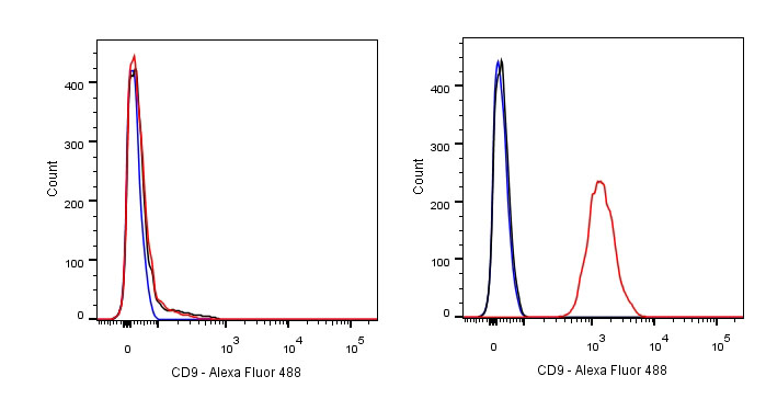
Flow cytometric analysis of Raji (left) / HCT 116 (right) cells labelling CD9 antibody at 1/500 (0.1ug) dilution/ (red) compared with a Rabbit monoclonal IgG (Black) isotype control and an unlabelled control (cells without incubation with primary antibody and secondary antibody) (Blue). Secondary antibody: #JP20025 was used as the secondary antibody. Negative control: Raji

CD9 Rabbit mAb at 1/25 dilution (2µg) immunoprecipitating CD9 in 0.4mg MCF7 whole cell lysate. Western blot was performed on the immunoprecipitate using CD9 Rabbit mAb at 1/1000 dilution. Secondary antibody (HRP) for IP was used at 1/400 dilution. Lane 1 : MCF7 whole cell lysate 10µg (input) Lane 2 : CD9 Rabbit mAb IP in MCF7 whole cell lysate Lane 3 : Rabbit monoclonal IgG IP in MCF7 whole cell lysate Predicted MW: 22 kDa Observed MW: 22 kDa Exposure time: 80s