
Mouse anti-PPARA Monoclonal Antibody(1331CT894.186.143)描述别名宿主特异性反应种属应用分子量类型克隆号同种型储存/保存方法研究领域背景说明细胞定位UniProt参考文献
| 概述 | |
| 描述 |
Purified Mouse Monoclonal Antibody (Mab)
|
| 别名 |
PPARA抗体;Peroxisome proliferator-activated receptor alpha; PPAR-alpha; Nuclear receptor subfamily 1 group C member 1; PPARA; NR1C1; PPAR
|
| 宿主 |
Mouse
|
| 特异性 |
This PPARA antibody is generated from a mouse immunized with a recombination protein from the human region of human PPARA.
|
| 反应种属 |
Human, Mouse
|
| 应用 |
FC~~1:25
IF~~1:25 IHC-P~~1:25 WB~~1:1000 |
| 分子量 |
Predicted molecular weight: 52kD
Disclaimer note: The observed molecular weight of the protein may vary from the listed predicted molecular weight due to post translational modifications, post translation cleavages, relative charges, and other experimental factors. |
| 性能 | |
| 类型 |
Monoclonal Antibody
|
| 克隆号 |
1331CT894.186.143
|
| 同种型 |
IgG1,κ
|
| 储存/保存方法 |
Maintain refrigerated at 2-8°C for up to 2 weeks. For long time storage store at -20°C in small aliquots to prevent freeze-thaw cycles.
|
| 研究领域 |
Cancer;Cardiovascular;Metabolism
|
| 靶标 | |
| 背景说明 |
Ligand-activated transcription factor. Key regulator of lipid metabolism. Activated by the endogenous ligand 1-palmitoyl- 2-oleoyl-sn-glycerol-3-phosphocholine (16:0/18:1-GPC). Activated by oleylethanolamide, a naturally occurring lipid that regulates satiety. Receptor for peroxisome proliferators such as hypolipidemic drugs and fatty acids. Regulates the peroxisomal beta-oxidation pathway of fatty acids. Functions as transcription activator for the ACOX1 and P450 genes. Transactivation activity requires heterodimerization with RXRA and is antagonized by NR2C2. May be required for the propagation of clock information to metabolic pathways regulated by PER2.
|
| 细胞定位 |
Nucleus.
|
| UniProt |
Q07869
|
| 参考文献 | |
| 参考文献 |
Sher T.,et al.Biochemistry 32:5598-5604(1993).
Mukherjee R.,et al.J. Steroid Biochem. Mol. Biol. 51:157-166(1994). Tugwood J.D.,et al.Ann. N. Y. Acad. Sci. 804:252-265(1996). Kobayashi T.,et al.FEBS Lett. 582:2737-2744(2008). Cho M.-C.,et al.Immunopharmacol. Immunotoxicol. 31:459-467(2009). |
实验结果图
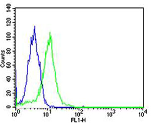
Flow cytometric analysis of Hela cells using PPARA Antibody(green, Cat#JP100453) compared to an isotype control of mouse IgG1(blue). JP100453 was diluted at 1:25 dilution. An Alexa Fluor® 488 goat anti-mouse lgG at 1:400 dilution was used as the secondary antibody.

Fluorescent image of Hela cells stained with PPARA Antibody(Cat#JP100453). JP100453 was diluted at 1:25 dilution. An Alexa Fluor® 488-conjugated goat anti-mouse lgG at 1:400 dilution was used as the secondary antibody (green). Cytoplasmic actin was counterstained with Alexa Fluor® 555 conjugated with Phalloidin (red).

Immunohistochemical analysis of paraffin-embedded H. skeletal muscle section using PPARA Antibody(Cat#JP100453). JP100453 was diluted at 1:25 dilution. A peroxidase-conjugated goat anti-mouse IgG at 1:400 dilution was used as the secondary antibody, followed by DAB staining.

Immunohistochemical analysis of paraffin-embedded H. kidney section using PPARA Antibody(Cat#JP100453). JP100453 was diluted at 1:25 dilution. A peroxidase-conjugated goat anti-mouse IgG at 1:400 dilution was used as the secondary antibody, followed by DAB staining.

Western blot analysis of lysates from Hela, Jurkat, mouse NIH/3T3 cell line (from left to right), using PPARA Antibody(Cat. #JP100453). JP100453 was diluted at 1:1000 at each lane. A goat anti-mouse IgG H&L(HRP) at 1:3000 dilution was used as the secondary antibody. Lysates at 35μg per lane.