
Rabbit anti-PCNA Recombinant Monoclonal Antibody(297-116)描述别名宿主特异性反应种属应用免疫原形式浓度纯化方法类型克隆号储存/保存方法存储溶液背景说明翻译后修饰细胞定位UniProt
| 概述 | |
| 描述 |
Proliferating cell nuclear antigen (PCNA) is a member of the DNA sliding clamp family of proteins that assist in DNA replication (1). Crystal structure data suggests that a PCNA homotrimer ring can encircle and slide along the DNA double helix (2). Multiple proteins involved in DNA replication, DNA repair, and cell cycle control bind to PCNA rather than directly associating with DNA, thus facilitating fast processing of DNA (reviewed in 3). PCNA protein expression is a well-accepted marker of proliferation and PCNA (PC10) Mouse mAb has been used to assess PCNA levels in hundreds of scientific studies.
|
| 别名 |
PCNA抗体;Proliferating cell nuclear antigen; Cyclin
|
| 宿主 |
Rabbit
|
| 特异性 |
PCNA Antibody detects endogenous levels of total PCNA.
|
| 反应种属 |
Human, Mouse, Rat
|
| 应用 |
WB: 1: 1000, IP: 1: 50, IHC-P: 1: 2000, ICC: 1: 500, FC(Intra): 1:50
|
| 免疫原 |
Synthetic peptide
|
| 性能 | |
| 形式 |
Liquid
|
| 浓度 |
0.5 mg/mL
|
| 纯化方法 |
Protein A affinity column
|
| 类型 |
Monoclonal Antibody
|
| 克隆号 |
297-116
|
| 储存/保存方法 |
Store at -20℃ for one year.
|
| 存储溶液 |
PBS, 40% Glycerol, 0.05% BSA, 0.03% Proclin 300
|
| 靶标 | |
| 背景说明 |
Proliferating cell nuclear antigen (PCNA) is a type of DNA clamp that acts as a DNA polymerase in eukaryotic cells δ. The processing factor of is crucial for replication. PCNA is a homologous trimer that achieves its processability by surrounding DNA, where it serves as a scaffold for recruiting proteins involved in DNA replication, DNA repair, chromatin remodeling, and epigenetics. It is expressed in most tumors as a proliferative antigen.
|
| 翻译后修饰 |
Phosphorylated. Phosphorylation at Tyr-211 by EGFR stabilizes chromatin-associated PCNA.Acetylated by CREBBP and p300/EP300; preferentially acetylated by CREBBP on Lys-80, Lys-13 and Lys-14 and on Lys-77 by p300/EP300 upon loading on chromatin in response to UV irradiation (PubMed:24939902, PubMed:19419956). Lysine acetylation disrupts association with chromatin, hence promoting PCNA ubiquitination and proteasomal degradation in response to UV damage in a CREBBP- and EP300-dependent manner (PubMed:24939902). Acetylation disrupts interaction with NUDT15 and promotes degradation (PubMed:19419956).Ubiquitinated (PubMed:24939902, PubMed:20227374). Following DNA damage, can be either monoubiquitinated to stimulate direct bypass of DNA lesions by specialized DNA polymerases or polyubiquitinated to promote recombination-dependent DNA synthesis across DNA lesions by template switching mechanisms. Following induction of replication stress, monoubiquitinated by the UBE2B-RAD18 complex on Lys-164, leading to recruit translesion (TLS) polymerases, which are able to synthesize across DNA lesions in a potentially error-prone manner. An error-free pathway also exists and requires non-canonical polyubiquitination on Lys-164 through ‘Lys-63’ linkage of ubiquitin moieties by the E2 complex UBE2N-UBE2V2 and the E3 ligases, HLTF, RNF8 and SHPRH. This error-free pathway, also known as template switching, employs recombination mechanisms to synthesize across the lesion, using as a template the undamaged, newly synthesized strand of the sister chromatid. Monoubiquitination at Lys-164 also takes place in undamaged proliferating cells, and is mediated by the DCX(DTL) complex, leading to enhance PCNA-dependent translesion DNA synthesis. Sumoylated during S phase.Methylated on glutamate residues by ARMT1/C6orf211.
|
| 细胞定位 |
Nucleus
|
| UniProt |
P12004
|
实验结果图
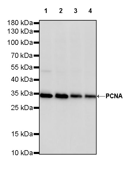
WB result of PCNA Rabbit mAb Primary antibody: PCNA Rabbit mAb at 1/1000 dilution Lane 1: HeLa whole cell lysate 20 ug Lane 2: HepG2 whole cell lysate 20 ug Lane 3: A431 whole cell lysate 20 ug Lane 4: MCF7 whole cell lysate 20 ug Secondary antibody: Goat Anti-Rabbit IgG, (H+L), HRP conjugated at 1/10000 dilution Predicted MW: 29 kDa Observed MW: 33 kDa Exposure time: 180 s
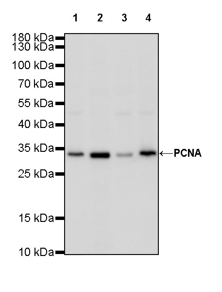
WB result of PCNA Rabbit mAb Primary antibody: PCNA Rabbit mAb at 1/1000 dilution Lane 1: mouse spleen lysate 20 ug Lane 2: mouse testis lysate 20 ug Lane 3: NIH/3T3 whole cell lysate 20 ug Lane 4: C2C12 whole cell lysate 20 ug Secondary antibody: Goat Anti-Rabbit IgG, (H+L), HRP conjugated at 1/10000 dilution Predicted MW: 29 kDa Observed MW: 33 kDa Exposure time: 180 s
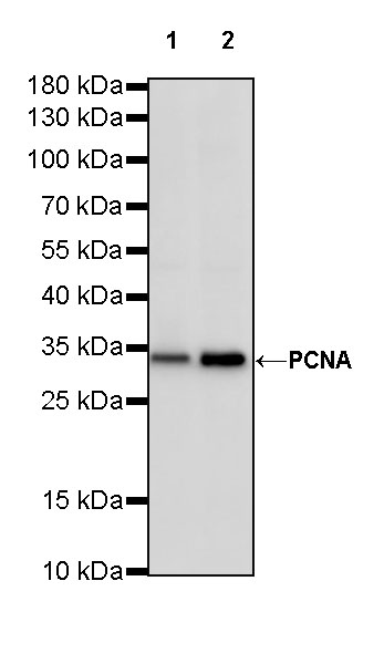
WB result of PCNA Rabbit mAb Primary antibody: PCNA Rabbit mAb at 1/1000 dilution Lane 1: PC-12 whole cell lysate 20 ug Lane 2: C6 whole cell lysate 20 ug Secondary antibody: Goat Anti-Rabbit IgG, (H+L), HRP conjugated at 1/10000 dilution Predicted MW: 29 kDa Observed MW: 33 kDa Exposure time: 180 s

PCNA Rabbit mAb at 1/50 dilution (1 ug) immunoprecipitating PCNA in 0.4 mg HeLa whole cell lysate. Western blot was performed on the immunoprecipitate using PCNA Rabbit mAb at 1/1000 dilution. Secondary antibody (HRP) for IP was used at 1/400 dilution. Lane 1: HeLa whole cell lysate 20 ug (Input) Lane 2: PCNA Rabbit mAb IP in HeLa whole cell lysate Lane 3: Rabbit monoclonal IgG IP in HeLa whole cell lysate Predicted MW: 29 kDa Observed MW: 33 kDa Exposure time: 30 s
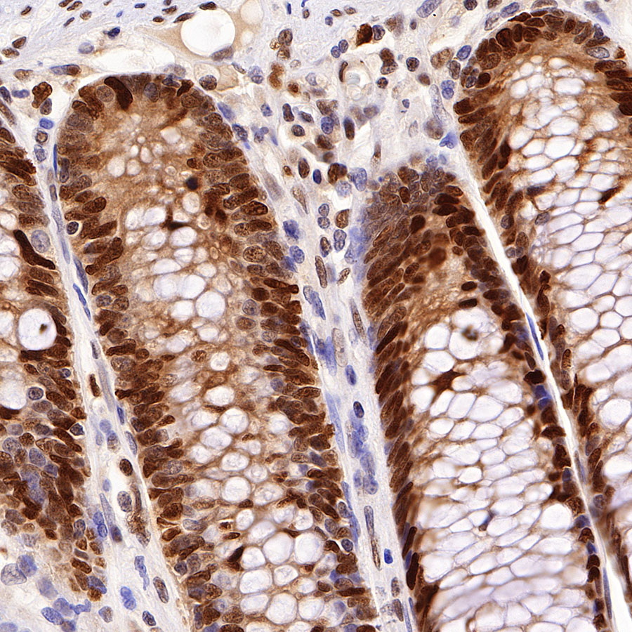
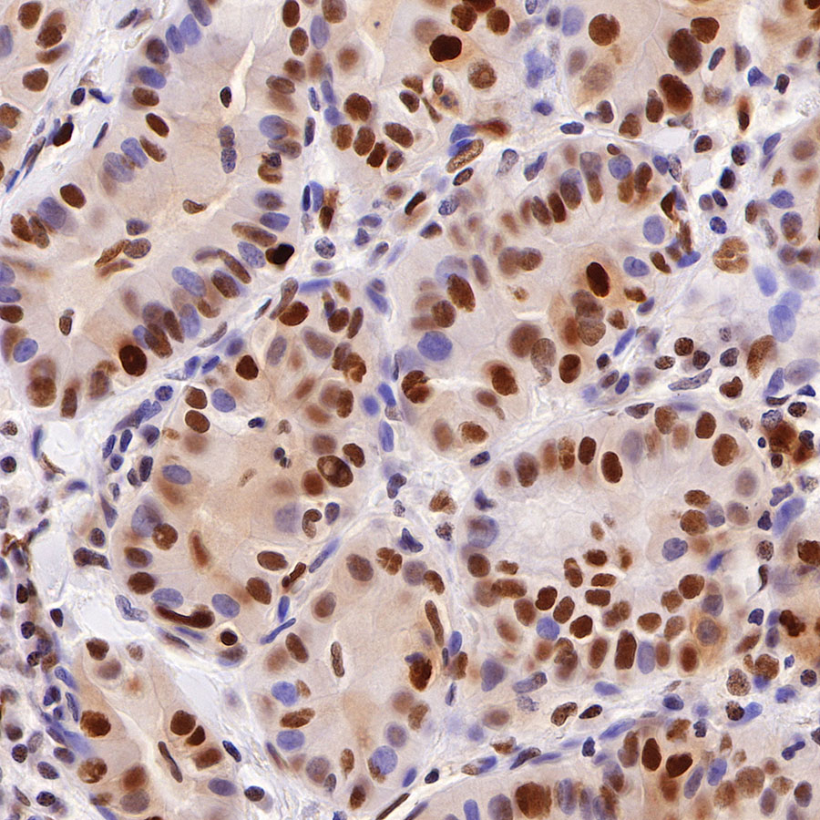
IHC shows positive staining in paraffin-embedded human colon. Anti-PCAN antibody was used at 1/2000 dilution, followed by a HRP Polymer for Mouse & Rabbit IgG (ready to use). Counterstained with hematoxylin. Heat mediated antigen retrieval with Tris/EDTA buffer pH9.0 was performed before commencing with IHC staining protocol.
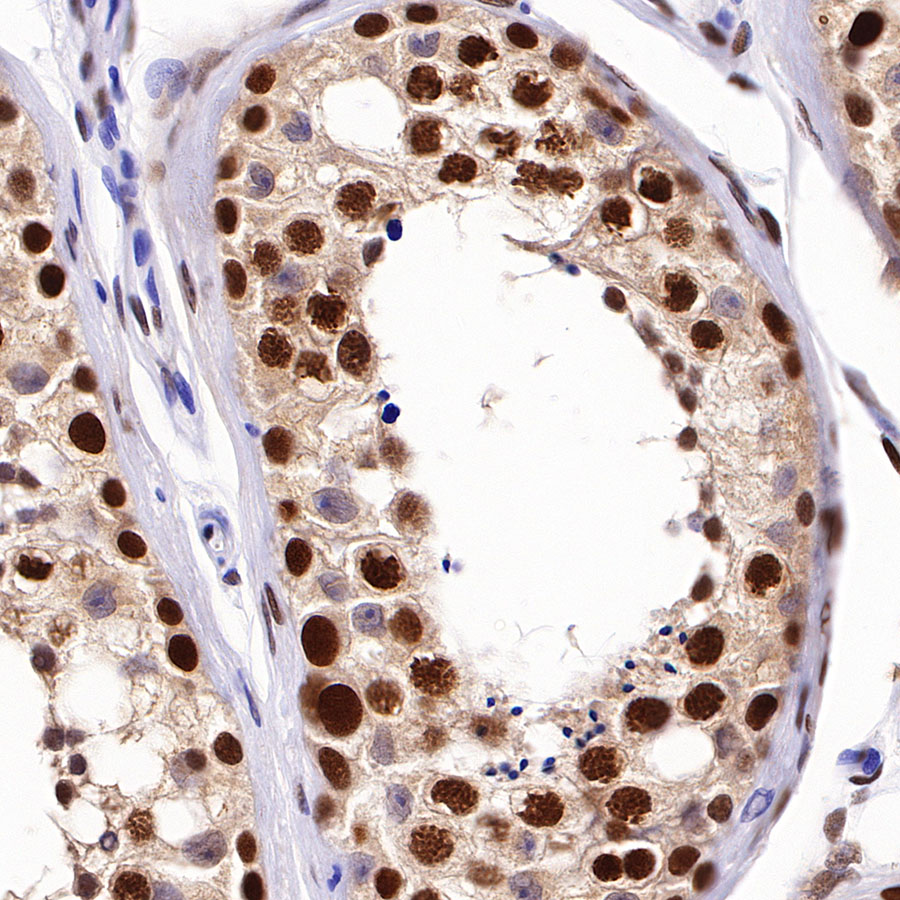
IHC shows positive staining in paraffin-embedded human testis. Anti-PCAN antibody was used at 1/2000 dilution, followed by a HRP Polymer for Mouse & Rabbit IgG (ready to use). Counterstained with hematoxylin. Heat mediated antigen retrieval with Tris/EDTA buffer pH9.0 was performed before commencing with IHC staining protocol.

IHC shows positive staining in paraffin-embedded human hepatocellular carcinoma. Anti-PCAN antibody was used at 1/2000 dilution, followed by a HRP Polymer for Mouse & Rabbit IgG (ready to use). Counterstained with hematoxylin. Heat mediated antigen retrieval with Tris/EDTA buffer pH9.0 was performed before commencing with IHC staining protocol.
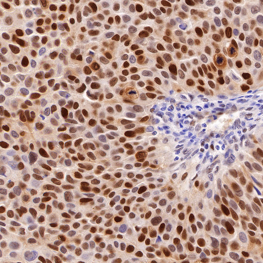
IHC shows positive staining in paraffin-embedded human bladder cancer. Anti-PCAN antibody was used at 1/2000 dilution, followed by a HRP Polymer for Mouse & Rabbit IgG (ready to use). Counterstained with hematoxylin. Heat mediated antigen retrieval with Tris/EDTA buffer pH9.0 was performed before commencing with IHC staining protocol.
IHC shows positive staining in paraffin-embedded human thyroid cancer. Anti-PCAN antibody was used at 1/2000 dilution, followed by a HRP Polymer for Mouse & Rabbit IgG (ready to use). Counterstained with hematoxylin. Heat mediated antigen retrieval with Tris/EDTA buffer pH9.0 was performed before commencing with IHC staining protocol.

IHC shows positive staining in paraffin-embedded mouse colon. Anti-PCAN antibody was used at 1/2000 dilution, followed by a HRP Polymer for Mouse & Rabbit IgG (ready to use). Counterstained with hematoxylin. Heat mediated antigen retrieval with Tris/EDTA buffer pH9.0 was performed before commencing with IHC staining protocol.
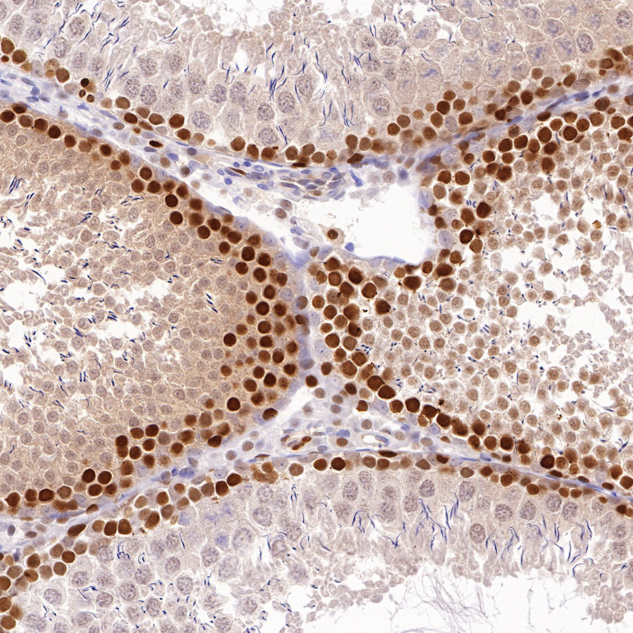
IHC shows positive staining in paraffin-embedded rat testis. Anti-PCAN antibody was used at 1/2000 dilution, followed by a HRP Polymer for Mouse & Rabbit IgG (ready to use). Counterstained with hematoxylin. Heat mediated antigen retrieval with Tris/EDTA buffer pH9.0 was performed before commencing with IHC staining protocol.
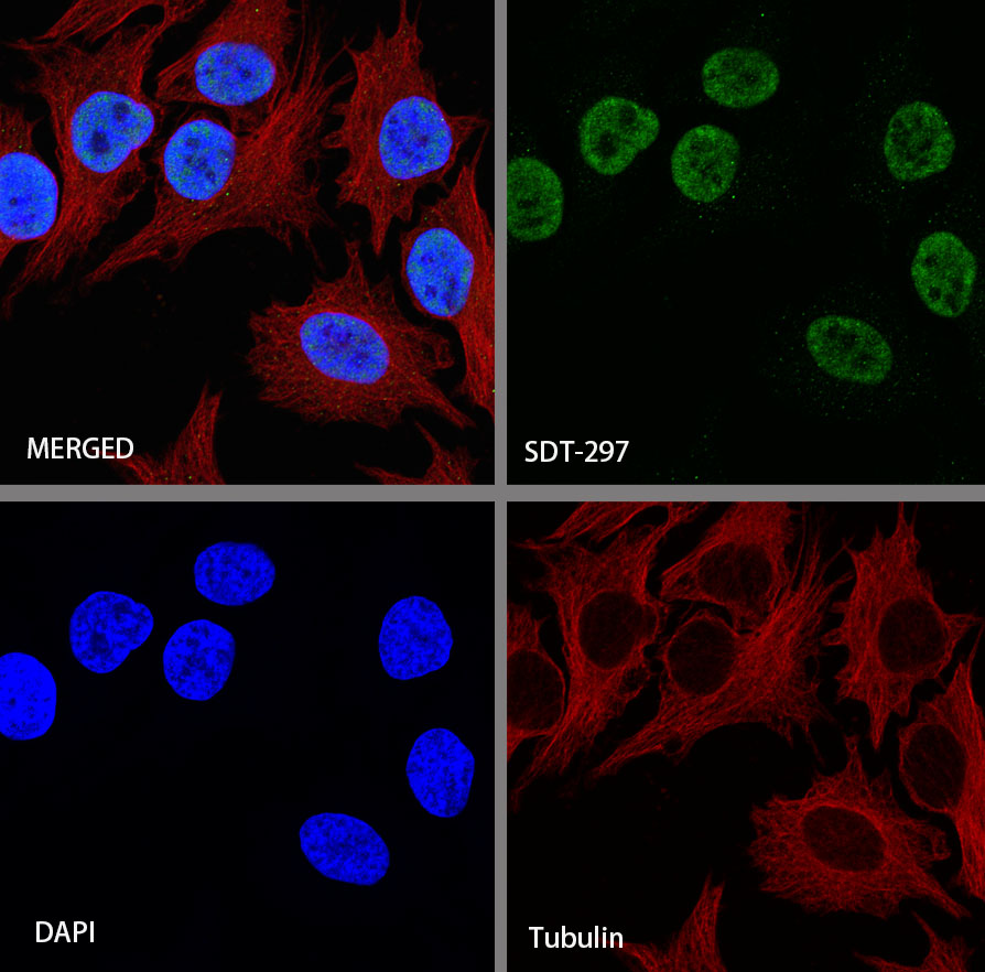
ICC shows positive staining in HeLa cells. Anti-PCNA antibody was used at 1/500 dilution (Green) and incubated overnight at 4°C. Goat polyclonal Antibody to Rabbit IgG – H&L (Alexa Fluor® 488) was used as secondary antibody at 1/1000 dilution. The cells were fixed with 4% PFA and permeabilized with 0.1% PBS-Triton X-100. Nuclei were counterstained with DAPI (Blue). Counterstain with tubulin (red).
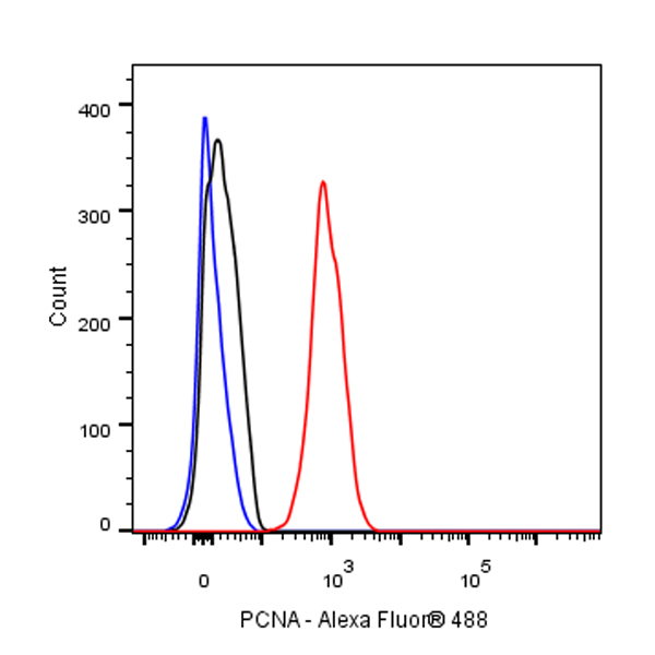
Flow cytometric analysis of 4% PFA fixed 90% methanol permeabilized HeLa (Human cervix adenocarcinoma epithelial cell) cells labelling PCNA antibody at 1/50 dilution (1 μg) / (red) compared with a Rabbit monoclonal IgG (Black) isotype control and an unlabelled control (cells without incubation with primary antibody and secondary antibody) (Blue). Goat Anti – Rabbit IgG Alexa Fluor® 488 was used as the secondary antibody.