
Mouse anti-VCP Monoclonal Antibody(1344CT150.163.114)描述别名宿主特异性反应种属应用分子量纯度类型克隆号同种型储存/保存方法研究领域背景说明细胞定位UniProt参考文献
| 概述 | |
| 描述 |
Purified Mouse Monoclonal Antibody (Mab)
|
| 别名 |
VCP抗体;Transitional endoplasmic reticulum ATPase; TER ATPase; 15S Mg(2+)-ATPase p97 subunit; Valosin-containing protein; VCP
|
| 宿主 |
Mouse
|
| 特异性 |
This VCP antibody is generated from a mouse immunized with a recombinant protein from human VCP.
|
| 反应种属 |
Human, Mouse, Rat
|
| 应用 |
IF~~1:50
WB~~1:2000 |
| 分子量 |
Predicted molecular weight: 89kD
Disclaimer note: The observed molecular weight of the protein may vary from the listed predicted molecular weight due to post translational modifications, post translation cleavages, relative charges, and other experimental factors. |
| 性能 | |
| 纯度 |
Greater than 95% as determined by reducing SDS-PAGE.
|
| 类型 |
Monoclonal Antibody
|
| 克隆号 |
1344CT150.163.114
|
| 同种型 |
IgG1,κ
|
| 储存/保存方法 |
Maintain refrigerated at 2-8°C for up to 2 weeks. For long term storage store at -20°C in small aliquots to prevent freeze-thaw cycles.
|
| 研究领域 |
Metabolism;Neuroscience;Signal Transduction
|
| 靶标 | |
| 背景说明 |
Necessary for the fragmentation of Golgi stacks during mitosis and for their reassembly after mitosis. Involved in the formation of the transitional endoplasmic reticulum (tER). The transfer of membranes from the endoplasmic reticulum to the Golgi apparatus occurs via 50-70 nm transition vesicles which derive from part-rough, part-smooth transitional elements of the endoplasmic reticulum (tER). Vesicle budding from the tER is an ATP-dependent process. The ternary complex containing UFD1L, VCP and NPLOC4 binds ubiquitinated proteins and is necessary for the export of misfolded proteins from the ER to the cytoplasm, where they are degraded by the proteasome. The NPLOC4-UFD1L-VCP complex regulates spindle disassembly at the end of mitosis and is necessary for the formation of a closed nuclear envelope. Regulates E3 ubiquitin-protein ligase activity of RNF19A. Component of the VCP/p97-AMFR/gp78 complex that participates in the final step of the sterol-mediated ubiquitination and endoplasmic reticulum-associated degradation (ERAD) of HMGCR. Also involved in DNA damage response: recruited to double-strand breaks (DSBs) sites in a RNF8- and RNF168-dependent manner and promotes the recruitment of TP53BP1 at DNA damage sites. Recruited to stalled replication forks by SPRTN: may act by mediating extraction of DNA polymerase eta (POLH) to prevent excessive translesion DNA synthesis and limit the incidence of mutations induced by DNA damage. Required for cytoplasmic retrotranslocation of stressed/damaged mitochondrial outer-membrane proteins and their subsequent proteasomal degradation.
|
| 细胞定位 |
Cytoplasm, cytosol. Endoplasmic reticulum. Nucleus. Note=Present in the neuronal hyaline inclusion bodies specifically found in motor neurons from amyotrophic lateral sclerosis patients. Present in the Lewy bodies specifically found in neurons from Parkinson disease patients. Recruited to the cytoplasmic surface of the endoplasmic reticulum via interaction with AMFR/gp78. Following DNA double-strand breaks, recruited to the sites of damage. Recruited to stalled replication forks via interaction with SPRTN
|
| UniProt |
P55072
|
| 参考文献 | |
| 参考文献 |
Lamerdin J.E.,et al.Submitted (MAR-1998) to the EMBL/GenBank/DDBJ databases.
Hu R.-M.,et al.Proc. Natl. Acad. Sci. U.S.A. 97:9543-9548(2000). Ota T.,et al.Nat. Genet. 36:40-45(2004). Humphray S.J.,et al.Nature 429:369-374(2004). Mural R.J.,et al.Submitted (SEP-2005) to the EMBL/GenBank/DDBJ databases. |
实验结果图
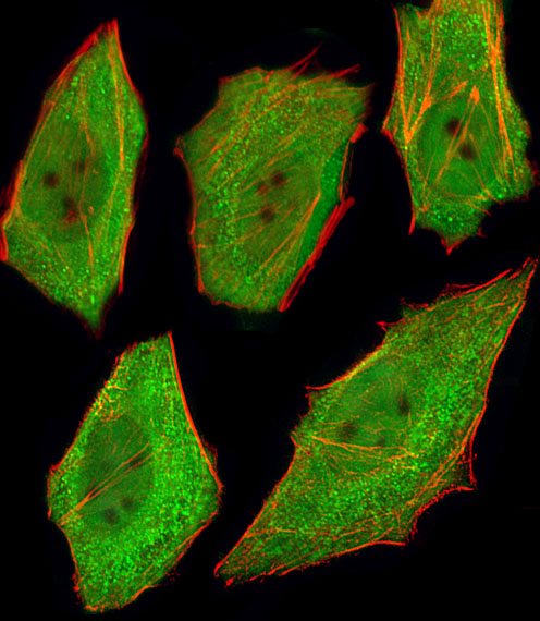
Fluorescent image of U251 cells stained with VCP Antibody (Cat#JP100416). JP100416 was diluted at 1:25 dilution. An Alexa Fluor® 488-conjugated goat anti-mouse lgG at 1:400 dilution was used as the secondary antibody (green). Cytoplasmic actin was counterstained with Alexa Fluor® 555 conjugated with Phalloidin (red).
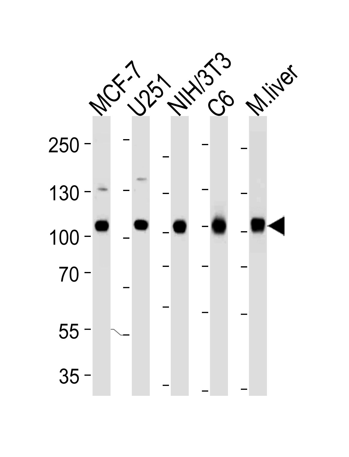
Western blot analysis of lysates from MCF-7, U251, mouse NIH/3T3, rat C6 cell line and mouse liver tissue lysate (from left to right) using VCP Antibody (Cat. # JP100416). JP100416 was diluted at 1:1000 at each lane. A goat anti-mouse IgG H&L(HRP) at 1:3000 dilution was used as the secondary antibody. Lysates at 35μg per lane.