
Rabbit anti-CytokeRatin 19 Recombiant Monoclonal Antibody(107-19)描述别名宿主特异性反应种属应用免疫原形式浓度纯化方法类型克隆号储存/保存方法存储溶液背景说明组织特异性细胞定位UniProt
| 概述 | |
| 描述 |
Expressed in a defined zone of basal keratinocytes in the deep outer root sheath of hair follicles. Also observed in sweat gland and mammary gland ductal and secretory cells, bile ducts, gastrointestinal tract, bladder urothelium, oral epithelia, esophagus, ectocervical epithelium (at protein level). Expressed in epidermal basal cells, in nipple epidermis and a defined region of the hair follicle. Also seen in a subset of vascular wall cells in both the veins and artery of human umbilical cord, and in umbilical cord vascular smooth muscle. Observed in muscle fibers accumulating in the costameres of myoplasm at the sarcolemma in structures that contain dystrophin and spectrin
|
| 别名 |
Cytokeratin 19抗体;KRT19; CK-19
|
| 宿主 |
Rabbit
|
| 特异性 |
Cytokeratin 19 Antibody detects endogenous levels of total Cytokeratin 19.
|
| 反应种属 |
Human, Mouse, Rat
|
| 应用 |
WB: 1:500, IHC:1:1000, FC: 1:500
|
| 免疫原 |
Synthetic peptide
|
| 性能 | |
| 形式 |
liquid
|
| 浓度 |
0.25mg/ml
|
| 纯化方法 |
Protein A affinity column
|
| 类型 |
Monoclonal antibody
|
| 克隆号 |
107-19
|
| 储存/保存方法 |
Store at -20℃ for one year.
|
| 存储溶液 |
PBS, 40% Glycerol, 0.05% BSA, 0.03% Proclin 300
|
| 靶标 | |
| 背景说明 |
Cytokeratin 19 is a member of the keratin family. The keratins are intermediate filament proteins responsible for the structural integrity of epithelial cells and are subdivided into cytokeratins and hair keratins. Keratin 19 is a type I keratin. The type I cytokeratins consist of acidic proteins which are arranged in pairs of heterotypic keratin chains. Unlike its related family members, this smallest known acidic cytokeratin is not paired with a basic cytokeratin in epithelial cells. It is specifically found in the periderm, the transiently superficial layer that envelops the developing epidermis. KRT19 is also known as Cyfra 21-1.Due to its high sensitivity, KRT19 is the most used marker for the RT-PCR-mediated detection of tumor cells disseminated in lymph nodes, peripheral blood, and bone marrow of breast cancer patients.
|
| 组织特异性 |
Expressed in a defined zone of basal keratinocytes in the deep outer root sheath of hair follicles. Also observed in sweat gland and mammary gland ductal and secretory cells, bile ducts, gastrointestinal tract, bladder urothelium, oral epithelia, esophagus, ectocervical epithelium (at protein level). Expressed in epidermal basal cells, in nipple epidermis and a defined region of the hair follicle. Also seen in a subset of vascular wall cells in both the veins and artery of human umbilical cord, and in umbilical cord vascular smooth muscle. Observed in muscle fibers accumulating in the costameres of myoplasm at the sarcolemma in structures that contain dystrophin and spectrin.
|
| 细胞定位 |
Cell membrane
|
| UniProt |
P08727
|
实验结果图
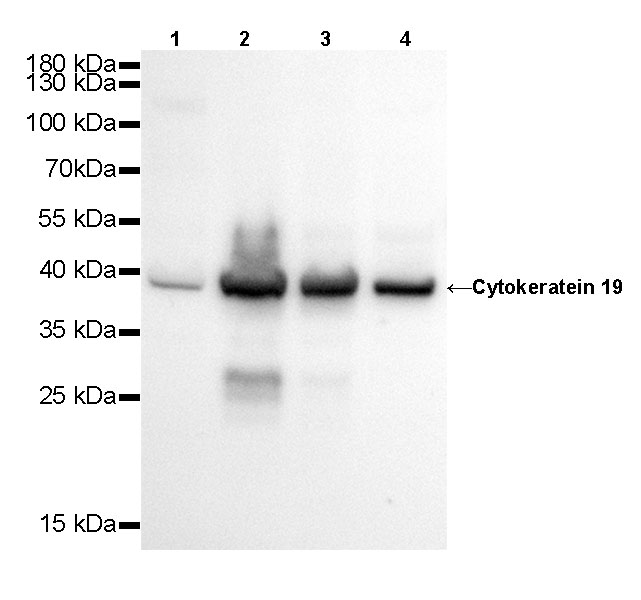
WB result of Cytokeratin 19 Rabbit mAb Primary antibody: Cytokeratin 19 Rabbit mAb at 1/500 dilution Lane 1: Hela whole cell lysate 20 µg Lane 2: MCF7 whole cell lysate 20 µg Lane 3: HT-29 whole cell lysate 20 µg Lane 4: HepG2 whole cell lysate 20 µgSecondary antibody: #JP20040 at 1/10000 dilution Predicted MW: 41 kDa Observed MW: 39 kDa Exposure time: 20s
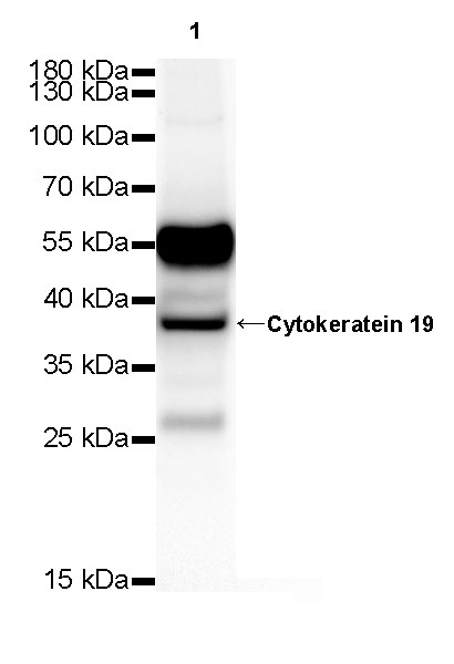
WB result of Cytokeratin 19 Rabbit mAb Primary antibody: Cytokeratin 19 Rabbit mAb at 1/500 dilution Lane 1: mouse kidney lysate 20 µg Secondary antibody: #JP20040 at 1/10000 dilution Predicted MW: 41 kDa Observed MW: 39 kDa Exposure time: 120 s
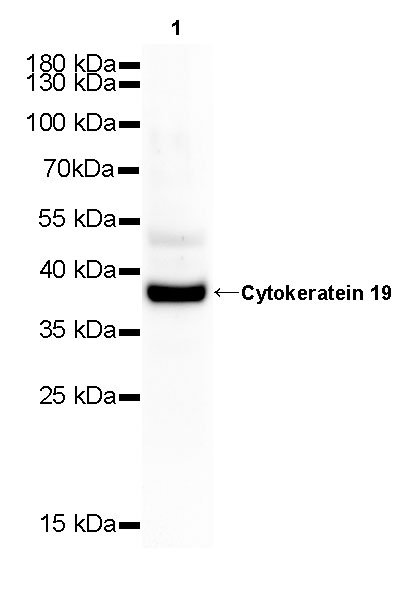
WB result of Cytokeratin 19 Rabbit mAb Primary antibody: Cytokeratin 19 Rabbit mAb at 1/500 dilution Lane 1: rat kidney whole cell lysate 20 µg Secondary antibody: #JP20040 at 1/10000 dilution Predicted MW: 41 kDa Observed MW: 39 kDa Exposure time: 120 s
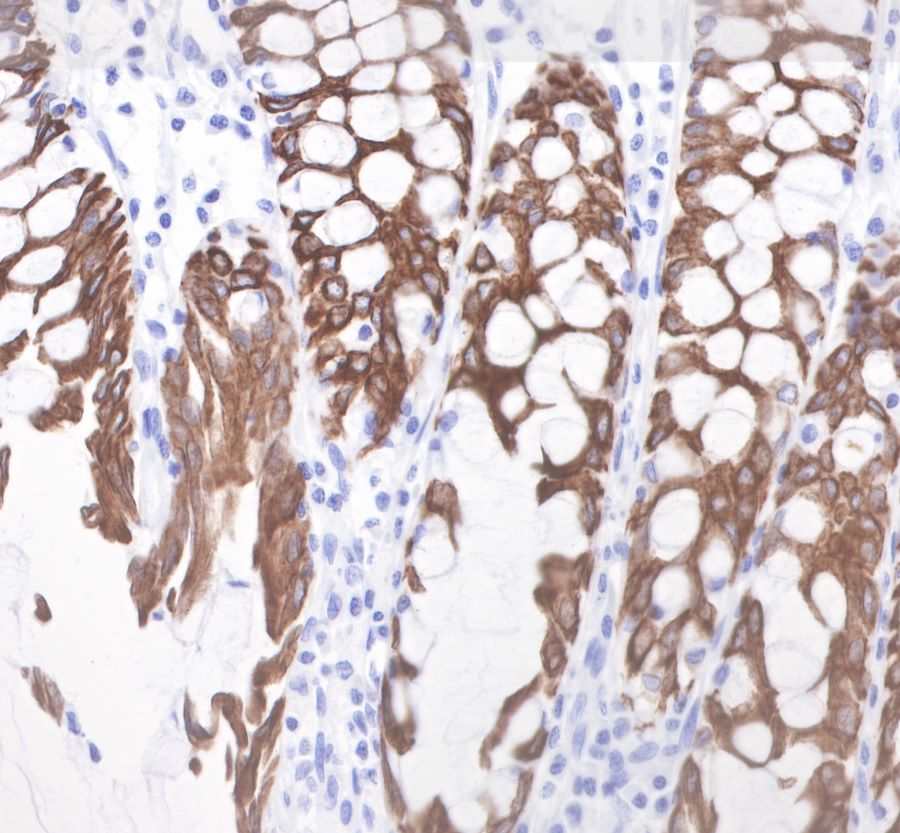
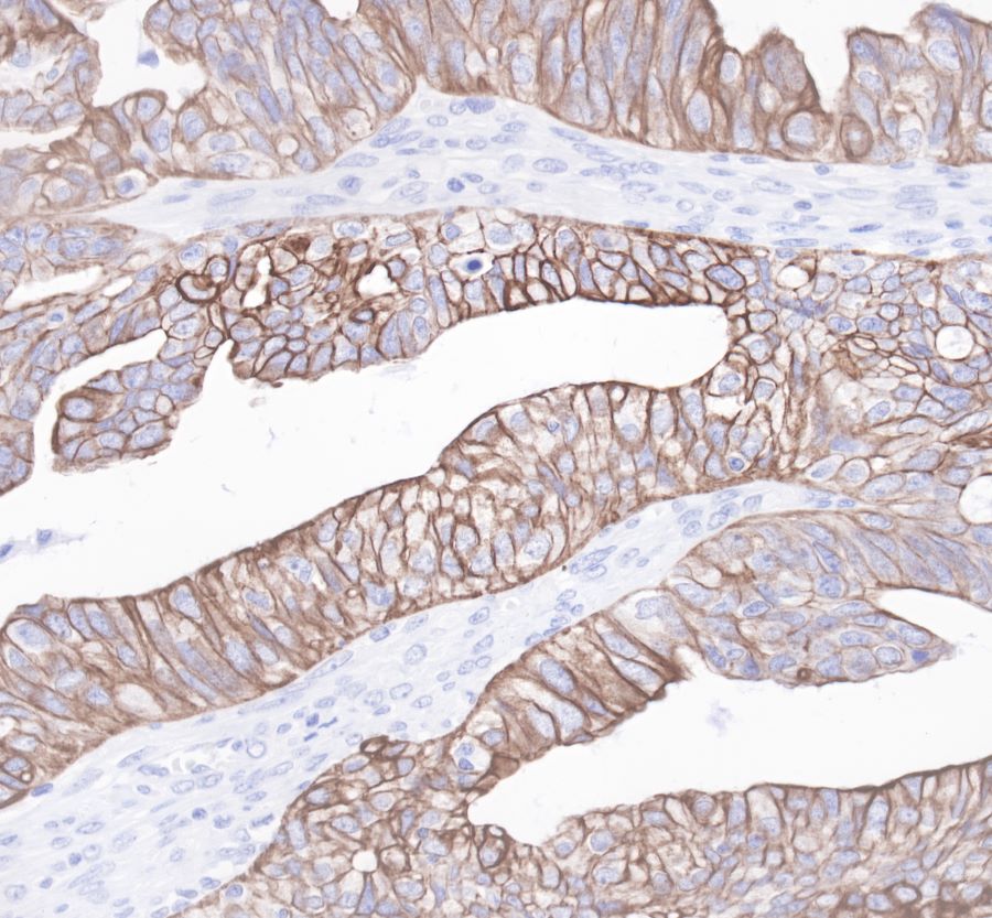
IHC shows positive staining in paraffin-embedded human colon. Anti-Cytokeratin19 antibody was used at 1/1000 dilution, Secondary antibody: #JP20040. Counterstained with hematoxylin. Heat mediated antigen retrieval with Tris/EDTA buffer pH9.0 was performed before commencing with IHC staining protocol.
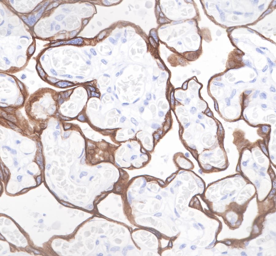
IHC shows positive staining in paraffin-embedded human placenta. Anti-Cytokeratin19 antibody was used at 1/1000 dilution, Secondary antibody: #JP20040. Counterstained with hematoxylin. Heat mediated antigen retrieval with Tris/EDTA buffer pH9.0 was performed before commencing with IHC staining protocol.
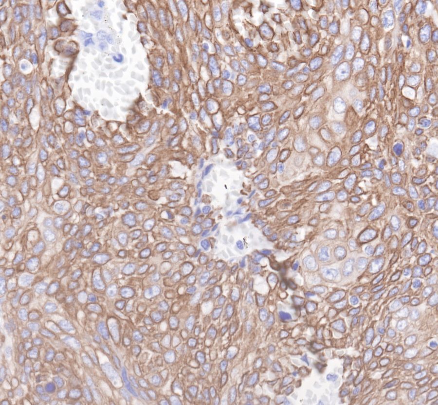
IHC shows positive staining in paraffin-embedded human ovarian cancer. Anti-Cytokeratin19 antibody was used at 1/1000 dilution, Secondary antibody: #JP20040. Counterstained with hematoxylin. Heat mediated antigen retrieval with Tris/EDTA buffer pH9.0 was performed before commencing with IHC staining protocol.
IHC shows positive staining in paraffin-embedded human cervix cancer. Anti-Cytokeratin19 antibody was used at 1/1000 dilution, Secondary antibody: #JP20040. Counterstained with hematoxylin. Heat mediated antigen retrieval with Tris/EDTA buffer pH9.0 was performed before commencing with IHC staining protocol.
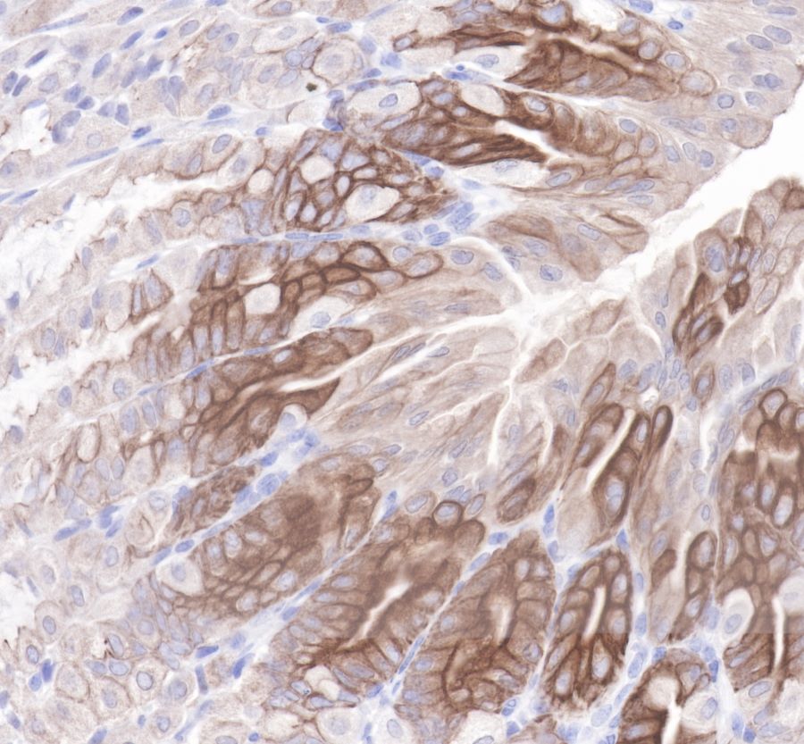
IHC shows positive staining in paraffin-embedded mouse stomach. Anti-Cytokeratin19 antibody was used at 1/1000 dilution, Secondary antibody: #JP20040. Counterstained with hematoxylin. Heat mediated antigen retrieval with Tris/EDTA buffer pH9.0 was performed before commencing with IHC staining protocol.
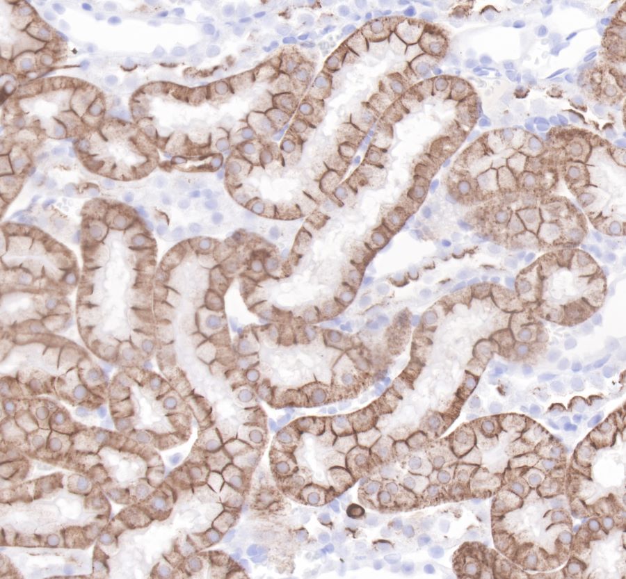
IHC shows positive staining in paraffin-embedded rat kidney. Anti-Cytokeratin19 antibody was used at 1/1000 dilution, Secondary antibody: #JP20040. Counterstained with hematoxylin. Heat mediated antigen retrieval with Tris/EDTA buffer pH9.0 was performed before commencing with IHC staining protocol.
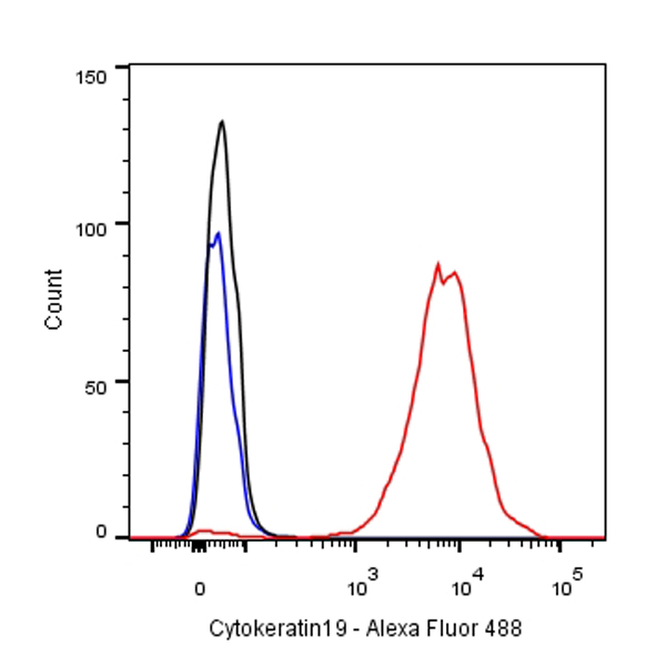
Flow cytometric analysis of MCF7 cells labelling Cytokeratin 19 antibody at 1/500 dilution/ (red) compared with a Rabbit monoclonal IgG (Black) isotype control and an unlabelled control (cells without incubation with primary antibody and secondary antibody) (Blue). Secondary antibody: #JP20040 at 1/1000 dilution was used as the secondary antibody.