
Mouse anti-RAC1 Monoclonal Antibody(1301CT276.121.104)描述别名宿主特异性反应种属预测反应种属应用分子量类型克隆号同种型储存/保存方法研究领域背景说明细胞定位UniProt参考文献
| 概述 | |
| 描述 |
Purified Mouse Monoclonal Antibody (Mab)
|
| 别名 |
RAC1抗体;Ras-related C3 botulinum toxin substrate 1; Cell migration-inducing gene 5 protein; Ras-like protein TC25; p21-Rac1; RAC1; TC25
|
| 宿主 |
Mouse
|
| 特异性 |
This antibody is generated from a mouse immunized with a KLH conjugated synthetic peptide between amino acids from human.
|
| 反应种属 |
Human, Rat
|
| 预测反应种属 |
Bovine, Mouse
|
| 应用 |
IHC-P~~1:25
FC~~1:100 WB~~1:1000 |
| 分子量 |
Predicted molecular weight: 21kD
Disclaimer note: The observed molecular weight of the protein may vary from the listed predicted molecular weight due to post translational modifications, post translation cleavages, relative charges, and other experimental factors. |
| 性能 | |
| 类型 |
Monoclonal Antibody
|
| 克隆号 |
1301CT276.121.104
|
| 同种型 |
IgG2b
|
| 储存/保存方法 |
Maintain refrigerated at 2-8°C for up to 2 weeks. For long term storage store at -20°C in small aliquots to prevent freeze-thaw cycles.
|
| 研究领域 |
Cancer;Cell Biology;Immunology;Signal Transduction;Amyotrophic Lateral Sclerosis ALS
|
| 靶标 | |
| 背景说明 |
Plasma membrane-associated small GTPase which cycles between active GTP-bound and inactive GDP-bound states. In its active state, binds to a variety of effector proteins to regulate cellular responses such as secretory processes, phagocytosis of apoptotic cells, epithelial cell polarization and growth-factor induced formation of membrane ruffles. Rac1 p21/rho GDI heterodimer is the active component of the cytosolic factor sigma 1, which is involved in stimulation of the NADPH oxidase activity in macrophages. Essential for the SPATA13-mediated regulation of cell migration and adhesion assembly and disassembly. Stimulates PKN2 kinase activity. In concert with RAB7A, plays a role in regulating the formation of RBs (ruffled borders) in osteoclasts. In glioma cells, promotes cell migration and invasion. In podocytes, promotes nuclear shuttling of NR3C2; this modulation is required for a proper kidney functioning. Required for atypical chemokine receptor ACKR2-induced LIMK1-PAK1-dependent phosphorylation of cofilin (CFL1) and for up-regulation of ACKR2 from endosomal compartment to cell membrane, increasing its efficiency in chemokine uptake and degradation. In synapses, seems to mediate the regulation of F-actin cluster formation performed by SHANK3.
|
| 细胞定位 |
Cell membrane; Lipid-anchor; Cytoplasmic side. Melanosome. Cytoplasm. Note=Inner surface of plasma membrane possibly with attachment requiring prenylation of the C-terminal cysteine (By similarity). Identified by mass spectrometry in melanosome fractions from stage I to stage IV. Found in the ruffled border (a late endosomal-like compartment in the plasma membrane) of bone-resorbing osteoclasts (By similarity).
|
| UniProt |
P63000
|
| 参考文献 | |
| 参考文献 |
Didsbury J.,et al.J. Biol. Chem. 264:16378-16382(1989).
Drivas G.T.,et al.Mol. Cell. Biol. 10:1793-1798(1990). Matos P.,et al.Biochem. Biophys. Res. Commun. 277:741-751(2000). Jordan P.,et al.Oncogene 18:6835-6839(1999). Schnelzer A.,et al.Submitted (MAR-1999) to the EMBL/GenBank/DDBJ databases. |
实验结果图
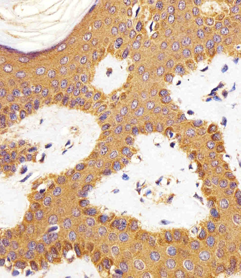
Immunohistochemical analysis of paraffin-embedded H.skin section using RAC1(Cat#JP100465). JP100465 was diluted at 1:25 dilution. A peroxidase-conjugated goat anti-mouse IgG at 1:400 dilution was used as the secondary antibody, followed by DAB staining.
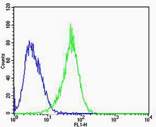
Flow cytometric analysis of U-87 MG cells using RAC1(green, Cat#JP100465) compared to an isotype control of mouse IgG2b(blue). AP20600c was diluted at 1:100 dilution. An Alexa Fluor® 488 goat anti-mouse lgG at 1:400 dilution was used as the secondary antibody.
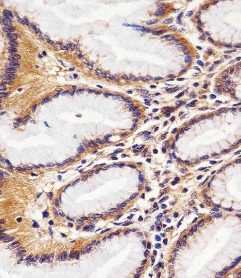
Immunohistochemical analysis of paraffin-embedded H.stomach section using RAC1(Cat#JP100465). JP100465 was diluted at 1:25 dilution. A peroxidase-conjugated goat anti-mouse IgG at 1:400 dilution was used as the secondary antibody, followed by DAB staining.
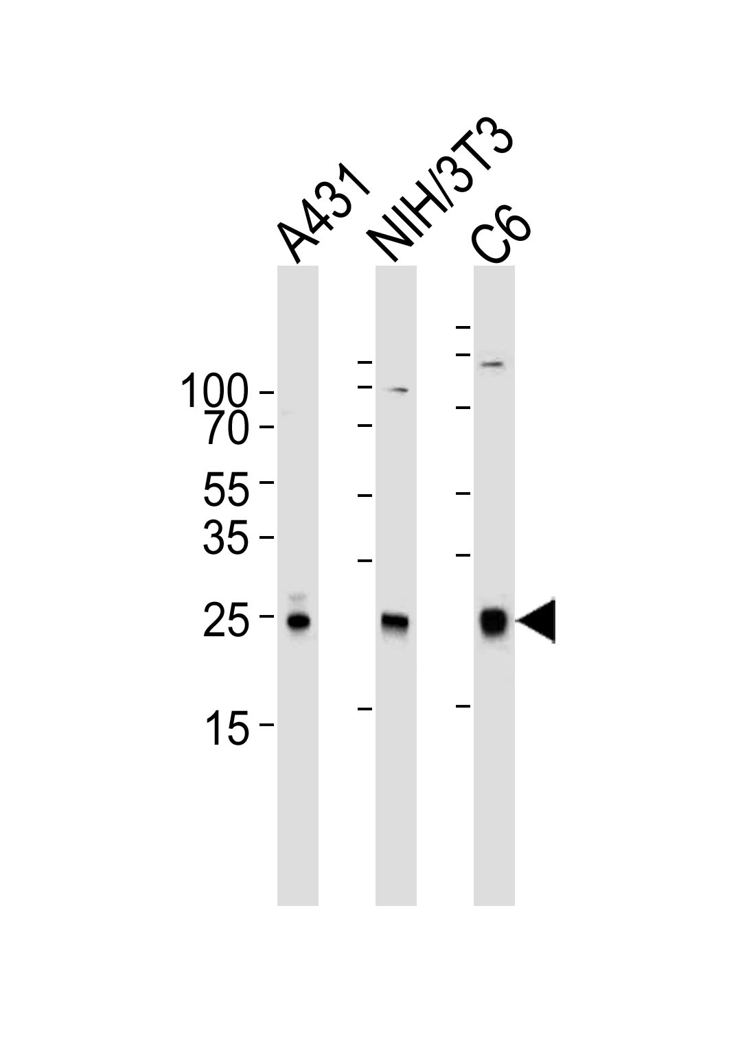
Western blot analysis of lysates from A431, mouse NIH/3T3, rat C6 cell line (from left to right), using RAC1 Antibody(Cat. #JP100465). JP100465 was diluted at 1:1000 at each lane. A goat anti-mouse IgG H&L(HRP) at 1:3000 dilution was used as the secondary antibody. Lysates at 35μg per lane.