
Mouse anti-PDIA1 Monoclonal Antibody(1530CT836.11.53)描述别名宿主特异性反应种属应用分子量类型克隆号同种型储存/保存方法研究领域背景说明细胞定位UniProt参考文献
| 概述 | |
| 描述 |
Purified Mouse Monoclonal Antibody (Mab)
|
| 别名 |
PDIA1抗体;Protein disulfide-isomerase; PDI; Cellular thyroid hormone-binding protein; Prolyl 4-hydroxylase subunit beta; p55; P4HB; ERBA2L; PDIA1; PO4DB
|
| 宿主 |
Mouse
|
| 特异性 |
This PDIA1 antibody is generated from a mouse immunized with a recombinant protein of human PDIA1.
|
| 反应种属 |
Human
|
| 应用 |
WB~~1:4000
|
| 分子量 |
Predicted molecular weight: 57kD
Disclaimer note: The observed molecular weight of the protein may vary from the listed predicted molecular weight due to post translational modifications, post translation cleavages, relative charges, and other experimental factors. |
| 性能 | |
| 类型 |
Monoclonal Antibody
|
| 克隆号 |
1530CT836.11.53
|
| 同种型 |
IgG1,k
|
| 储存/保存方法 |
Maintain refrigerated at 2-8°C for up to 2 weeks. For long term storage store at -20°C in small aliquots to prevent freeze-thaw cycles.
|
| 研究领域 |
Cancer;Metabolism;Signal Transduction
|
| 靶标 | |
| 背景说明 |
This multifunctional protein catalyzes the formation, breakage and rearrangement of disulfide bonds. At the cell surface, seems to act as a reductase that cleaves disulfide bonds of proteins attached to the cell. May therefore cause structural modifications of exofacial proteins. Inside the cell, seems to form/rearrange disulfide bonds of nascent proteins. At high concentrations, functions as a chaperone that inhibits aggregation of misfolded proteins. At low concentrations, facilitates aggregation (anti-chaperone activity). May be involved with other chaperones in the structural modification of the TG precursor in hormone biogenesis. Also acts a structural subunit of various enzymes such as prolyl 4-hydroxylase and microsomal triacylglycerol transfer protein MTTP.
|
| 细胞定位 |
Endoplasmic reticulum lumen. Melanosome. Cell membrane; Peripheral membrane protein. Note=Highly abundant. In some cell types, seems to be also secreted or associated with the plasma membrane, where it undergoes constant shedding and replacement from intracellular sources (Probable). Localizes near CD4-enriched regions on lymphoid cell surfaces. Identified by mass spectrometry in melanosome fractions from stage I to stage IV.
|
| UniProt |
P07237
|
| 参考文献 | |
| 参考文献 |
Pihlajaniemi T.,et al.EMBO J. 6:643-649(1987).
Cheng S.-Y.,et al.J. Biol. Chem. 262:11221-11227(1987). Tasanen K.,et al.J. Biol. Chem. 263:16218-16224(1988). Ota T.,et al.Nat. Genet. 36:40-45(2004). Mural R.J.,et al.Submitted (JUL-2005) to the EMBL/GenBank/DDBJ databases. |
实验结果图
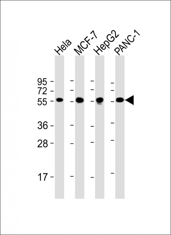
All lanes : Anti-PDIA1 Antibody at 1:4000 dilution Lane 1: Hela whole cell lysate Lane 2: MCF-7 whole cell lysate Lane 3: HepG2 whole cell lysate Lane 4: PANC-1 whole cell lysate Lysates/proteins at 20 µg per lane. Secondary Goat Anti-mouse IgG, (H+L), Peroxidase conjugated at 1/10000 dilution. Predicted band size : 57 kDa Blocking/Dilution buffer: 5% NFDM/TBST.