
Mouse anti-HINT1 Monoclonal Antibody(1500CT836.13.93)描述别名宿主特异性反应种属应用分子量类型克隆号同种型储存/保存方法研究领域背景说明细胞定位UniProt参考文献
| 概述 | |
| 描述 |
Purified Mouse Monoclonal Antibody (Mab)
|
| 别名 |
HINT1抗体;Histidine triad nucleotide-binding protein 1; 3—; Adenosine 5′-monophosphoramidase; Protein kinase C inhibitor 1; Protein kinase C-interacting protein 1; PKCI-1; HINT1; HINT; PKCI1; PRKCNH1
|
| 宿主 |
Mouse
|
| 特异性 |
This HINT1 antibody is generated from a mouse immunized with a recombinant protein of human HINT1.
|
| 反应种属 |
Human, Mouse, Rat
|
| 应用 |
IF~~1:25
IHC-P~~1:25 FC~~1:25 WB~~1:4000 |
| 分子量 |
Predicted molecular weight: 14kD
Disclaimer note: The observed molecular weight of the protein may vary from the listed predicted molecular weight due to post translational modifications, post translation cleavages, relative charges, and other experimental factors. |
| 性能 | |
| 类型 |
Monoclonal Antibody
|
| 克隆号 |
1500CT836.13.93
|
| 同种型 |
IgG1,k
|
| 储存/保存方法 |
Maintain refrigerated at 2-8°C for up to 2 weeks. For long term storage store at -20°C in small aliquots to prevent freeze-thaw cycles.
|
| 研究领域 |
Cancer
|
| 靶标 | |
| 背景说明 |
Hydrolyzes purine nucleotide phosphoramidates with a single phosphate group, including adenosine 5’monophosphoramidate (AMP-NH2), adenosine 5’monophosphomorpholidate (AMP-morpholidate) and guanosine 5’monophosphomorpholidate (GMP-morpholidate). Hydrolyzes lysyl-AMP (AMP-N-epsilon-(N-alpha-acetyl lysine methyl ester)) generated by lysine tRNA ligase, as well as Met-AMP, His- AMP and Asp-AMP, lysyl-GMP (GMP-N-epsilon-(N-alpha-acetyl lysine methyl ester)) and AMP-N-alanine methyl ester. Can also convert adenosine 5′-O-phosphorothioate and guanosine 5′-O- phosphorothioate to the corresponding nucleoside 5′-O-phosphates with concomitant release of hydrogen sulfide. In addition, functions as scaffolding protein that modulates transcriptional activation by the LEF1/TCF1-CTNNB1 complex and by the complex formed with MITF and CTNNB1. Modulates p53/TP53 levels and p53/TP53-mediated apoptosis. Modulates proteasomal degradation of target proteins by the SCF (SKP2-CUL1-F-box protein) E3 ubiquitin- protein ligase complex.
|
| 细胞定位 |
Cytoplasm. Nucleus. Note=Interaction with CDK7 leads to a more nuclear localization
|
| UniProt |
P49773
|
| 参考文献 | |
| 参考文献 |
Brzoska P.M.,et al.Genomics 36:151-156(1996).
Brzoska P.M.,et al.Proc. Natl. Acad. Sci. U.S.A. 92:7824-7828(1995). Ota T.,et al.Nat. Genet. 36:40-45(2004). Ebert L.,et al.Submitted (JUN-2004) to the EMBL/GenBank/DDBJ databases. Lima C.D.,et al.Proc. Natl. Acad. Sci. U.S.A. 93:5357-5362(1996). |
实验结果图
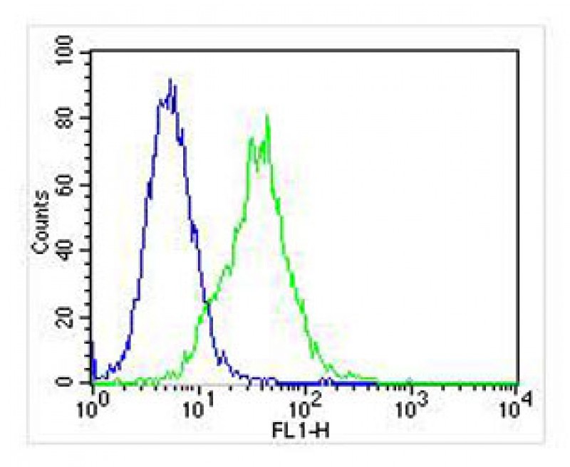
Overlay histogram showing Jurkat cells stained with JP100501 (green line). The cells were fixed with 2% paraformaldehyde (10 min) and then permeabilized with 90% methanol for 10 min. The cells were then icubated in 2% bovine serum albumin to block non-specific protein-protein interactions followed by the antibody (JP100501, 1:25 dilution) for 60 min at 37ºC. The secondary antibody used was Goat-Anti-Mouse IgG, DyLight® 488 Conjugated Highly Cross-Adsorbed(NA168821)) at 1/400 dilution for 40 min at 37ºC. Isotype control antibody (blue line) was mouse IgG1 (1μg/1×10^6 cells) used under the same conditions. Acquisition of >10, 000 events was performed.
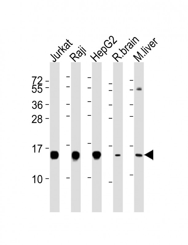
All lanes : Anti-HINT1 Antibody at 1:4000 dilution Lane 1: Jurkat whole cell lysates Lane 2: Raji whole cell lysates Lane 3: HepG2 whole cell lysates Lane 4: rat brain lysates Lane 5: mouse liver lysates Lysates/proteins at 20 μg per lane. Secondary Goat Anti-mouse IgG, (H+L), Peroxidase conjugated at 1/10000 dilution Predicted band size : 14 kDa Blocking/Dilution buffer: 5% NFDM/TBST.