
Mouse anti-VEGFR3 Monoclonal Antibody(818CT12.1.1)描述别名宿主特异性反应种属应用分子量类型克隆号同种型储存/保存方法存储溶液研究领域背景说明细胞定位UniProt参考文献
| 概述 | |
| 描述 |
Mouse Monoclonal Antibody (Mab)
|
| 别名 |
VEGFR3抗体;Vascular endothelial growth factor receptor 3; VEGFR-3; Fms-like tyrosine kinase 4; FLT-4; Tyrosine-protein kinase receptor FLT4; FLT4; VEGFR3
|
| 宿主 |
Mouse
|
| 特异性 |
Purified His-tagged VEGFR3 protein was used to produced this monoclonal antibody.
|
| 反应种属 |
Human
|
| 应用 |
FC~~1:25
IHC-P~~1:25 WB~~1:2000 |
| 分子量 |
Predicted molecular weight: 153kD
Disclaimer note: The observed molecular weight of the protein may vary from the listed predicted molecular weight due to post translational modifications, post translation cleavages, relative charges, and other experimental factors. |
| 性能 | |
| 类型 |
Monoclonal Antibody
|
| 克隆号 |
818CT12.1.1
|
| 同种型 |
IgG2a
|
| 储存/保存方法 |
Maintain refrigerated at 2-8°C for up to 2 weeks. For long term storage store at -20°C in small aliquots to prevent freeze-thaw cycles.
|
| 存储溶液 |
Purified monoclonal antibody supplied in PBS with 0.09% (W/V) sodium azide. This antibody is purified through a protein G column, eluted with high and low pH buffers and neutralized immediately, followed by dialysis against PBS.
|
| 研究领域 |
Cancer;Cardiovascular;Immunology;Signal Transduction
|
| 靶标 | |
| 背景说明 |
Tyrosine-protein kinase that acts as a cell-surface receptor for VEGFC and VEGFD, and plays an essential role in adult lymphangiogenesis and in the development of the vascular network and the cardiovascular system during embryonic development. Promotes proliferation, survival and migration of endothelial cells, and regulates angiogenic sprouting. Signaling by activated FLT4 leads to enhanced production of VEGFC, and to a lesser degree VEGFA, thereby creating a positive feedback loop that enhances FLT4 signaling. Modulates KDR signaling by forming heterodimers. The secreted isoform 3 may function as a decoy receptor for VEGFC and/or VEGFD and play an important role as a negative regulator of VEGFC-mediated lymphangiogenesis and angiogenesis. Binding of vascular growth factors to isoform 1 or isoform 2 leads to the activation of several signaling cascades; isoform 2 seems to be less efficient in signal transduction, because it has a truncated C-terminus and therefore lacks several phosphorylation sites. Mediates activation of the MAPK1/ERK2, MAPK3/ERK1 signaling pathway, of MAPK8 and the JUN signaling pathway, and of the AKT1 signaling pathway. Phosphorylates SHC1. Mediates phosphorylation of PIK3R1, the regulatory subunit of phosphatidylinositol 3- kinase. Promotes phosphorylation of MAPK8 at ‘Thr-183’ and ‘Tyr- 185’, and of AKT1 at ‘Ser-473’.
|
| 细胞定位 |
Cell membrane; Single-pass type I membrane protein. Cytoplasm. Nucleus. Note=Ligand-mediated autophosphorylation leads to rapid internalization Isoform 2: Cell membrane; Single-pass type I membrane protein
|
| UniProt |
P35916
|
| 参考文献 | |
| 参考文献 |
Irrthum A., et al. Am. J. Hum. Genet. 67:295-301(2000).
Pajusola K., et al. Cancer Res. 52:5738-5743(1992). Pajusola K., et al. Cancer Res. 53:3845-3845(1993). Galland F., et al. Genomics 13:475-478(1992). Galland F., et al. Oncogene 8:1233-1240(1993). |
实验结果图
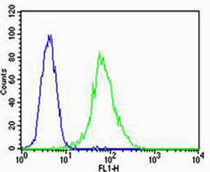
Flow cytometric analysis of HUVEC cells using VEGFR3(green, Cat#JP100388) compared to an isotype control of mouse IgG2a(blue). JP100388was diluted at 1:25 dilution. An Alexa Fluor® 488 goat anti-mouse lgG at 1:400 dilution was used as the secondary antibody.
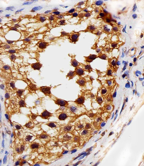
Immunohistochemical analysis of paraffin-embedded H. testis section using VEGFR3(Cat#JP100388). was diluted at 1:25 dilution. A peroxidase-conjugated goat anti-mouse IgG at 1:400 dilution was used as the secondary antibody, followed by DAB staining.
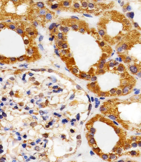
Immunohistochemical analysis of paraffin-embedded H. kidney section using VEGFR3(Cat#JP100388). was diluted at 1:25 dilution. A peroxidase-conjugated goat anti-mouse IgG at 1:400 dilution was used as the secondary antibody, followed by DAB staining.
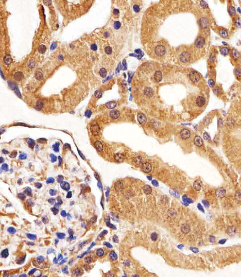
Immunohistochemical analysis of paraffin-embedded R. kidney section using VEGFR3(Cat#JP100388). NA was diluted at 1:25 dilution. A peroxidase-conjugated goat anti-mouse IgG at 1:400 dilution was used as the secondary antibody, followed by DAB staining.
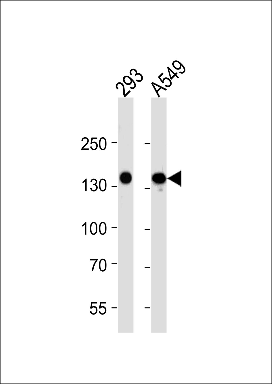
VEGFR3 Antibody (Cat. #JP100388) western blot analysis in 293 and A549 cell line lysates (35μg/lane).This demonstrates the VEGFR3 antibody detected the VEGFR3 protein (arrow).