
Mouse anti-ATG4A Monoclonal Antibody(1458CT808.66.25.69)描述别名宿主特异性反应种属应用分子量类型克隆号同种型储存/保存方法研究领域背景说明细胞定位UniProt参考文献
| 概述 | |
| 描述 |
Purified Mouse Monoclonal Antibody (Mab)
|
| 别名 |
ATG4A抗体;Cysteine protease ATG4A; 3422-; AUT-like 2 cysteine endopeptidase; Autophagin-2; Autophagy-related cysteine endopeptidase 2; Autophagy-related protein 4 homolog A; hAPG4A; ATG4A; APG4A; AUTL2
|
| 宿主 |
Mouse
|
| 特异性 |
This ATG4A antibody is generated from a mouse immunized with a recombinant protein.
|
| 反应种属 |
Human
|
| 应用 |
IF~~1:25
IHC-P~~1:25 FC~~1:25 WB~~1:500 |
| 分子量 |
Predicted molecular weight: 45kD
Disclaimer note: The observed molecular weight of the protein may vary from the listed predicted molecular weight due to post translational modifications, post translation cleavages, relative charges, and other experimental factors. |
| 性能 | |
| 类型 |
Monoclonal Antibody
|
| 克隆号 |
1458CT808.66.25.69
|
| 同种型 |
IgG2b,k
|
| 储存/保存方法 |
Maintain refrigerated at 2-8°C for up to 2 weeks. For long term storage store at -20°C in small aliquots to prevent freeze-thaw cycles.
|
| 研究领域 |
Cancer;Cardiovascular;Cell Biology;Metabolism
|
| 靶标 | |
| 背景说明 |
Cysteine protease required for the cytoplasm to vacuole transport (Cvt) and autophagy. Cleaves the C-terminal amino acid of ATG8 family proteins to reveal a C-terminal glycine. Exposure of the glycine at the C-terminus is essential for ATG8 proteins conjugation to phosphatidylethanolamine (PE) and insertion to membranes, which is necessary for autophagy. Preferred substrate is GABARAPL2 followed by MAP1LC3A and GABARAP. Has also an activity of delipidating enzyme for the PE-conjugated forms.
|
| 细胞定位 |
Cytoplasm.
|
| UniProt |
Q8WYN0
|
| 参考文献 | |
| 参考文献 |
Marino G.,et al.J. Biol. Chem. 278:3671-3678(2003).
Kabeya Y.,et al.J. Cell Sci. 117:2805-2812(2004). Chen J.M.,et al.Submitted (SEP-2001) to the EMBL/GenBank/DDBJ databases. Ota T.,et al.Nat. Genet. 36:40-45(2004). Ross M.T.,et al.Nature 434:325-337(2005). |
实验结果图
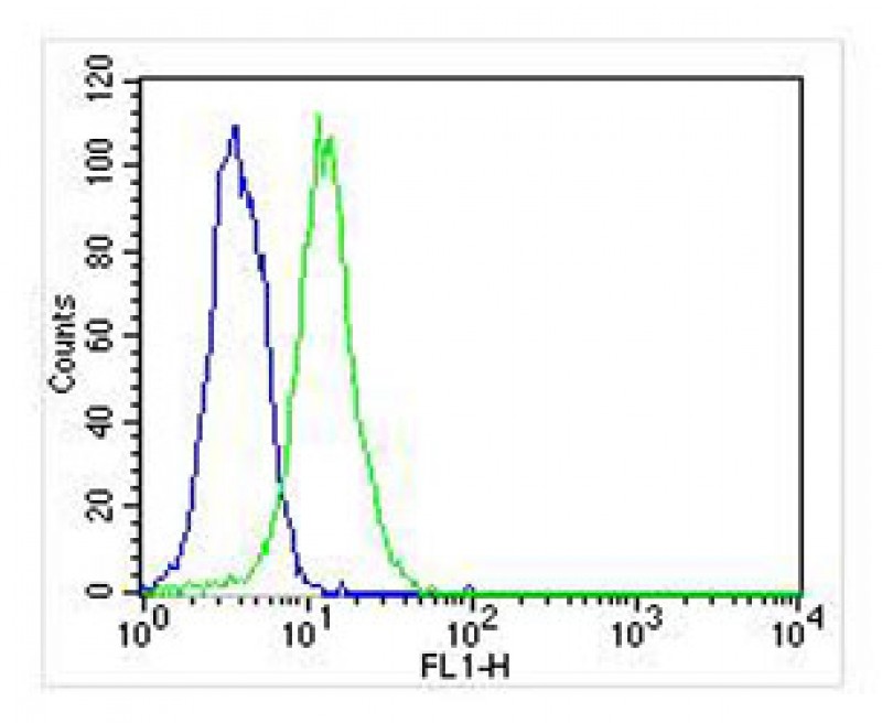
Overlay histogram showing Hela cells stained with JP100486 (green line). The cells were fixed with 2% paraformaldehyde (10 min) and then permeabilized with 90% methanol for 10 min. The cells were then icubated in 2% bovine serum albumin to block non-specific protein-protein interactions followed by the antibody (JP100486, 1:25 dilution) for 60 min at 37ºC. The secondary antibody used was Goat-Anti-Mouse IgG, DyLight® 488 Conjugated Highly Cross-Adsorbed(NA168821)) at 1/400 dilution for 40 min at 37ºC. Isotype control antibody (blue line) was mouse IgG2b (1μg/1×10^6 cells) used under the same conditions. Acquisition of >10, 000 events was performed.
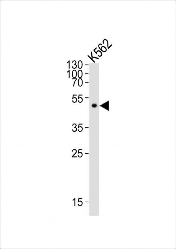
Western blot analysis of lysate from K562 cell line, using ATG4A Antibody(Cat. #JP100486). JP100486 was diluted at 1:500. A goat anti-mouse IgG H&L(HRP) at 1:10000 dilution was used as the secondary antibody. Lysate at 20μg.