
Rabbit anti-Tuberin Polyclonal Antibody(S1798)描述别名宿主特异性反应种属应用分子量类型同种型储存/保存方法存储溶液研究领域背景说明细胞定位UniProt参考文献
| 概述 | |
| 描述 |
Peptide Affinity Purified Rabbit Polyclonal Antibody (Pab)
|
| 别名 |
Tuberin抗体;Tuberin; Tuberous sclerosis 2 protein; TSC2; TSC4
|
| 宿主 |
Rabbit
|
| 特异性 |
This Tuberin (TSC2) antibody is generated from rabbits immunized with a KLH conjugated synthetic peptide between 1776-1805 amino acids from human Tuberin (TSC2).
|
| 反应种属 |
Human
|
| 应用 |
WB~~1:1000
IF~~1:10~50 IHC-P~~1:10~50 |
| 分子量 |
Predicted molecular weight: 201kD
Disclaimer note: The observed molecular weight of the protein may vary from the listed predicted molecular weight due to post translational modifications, post translation cleavages, relative charges, and other experimental factors. |
| 性能 | |
| 类型 |
Polyclonal Antibody
|
| 同种型 |
Rabbit Ig
|
| 储存/保存方法 |
Maintain refrigerated at 2-8°C for up to 2 weeks. For long term storage store at -20°C in small aliquots to prevent freeze-thaw cycles.
|
| 存储溶液 |
Purified polyclonal antibody supplied in PBS with 0.09% (W/V) sodium azide. This antibody is purified through a protein A column, followed by peptide affinity purification.
|
| 研究领域 |
Cell Biology;Metabolism;Signal Transduction
|
| 靶标 | |
| 背景说明 |
In complex with TSC1, inhibits the nutrient-mediated or growth factor-stimulated phosphorylation of S6K1 and EIF4EBP1 by negatively regulating mTORC1 signaling. Acts as a GTPase- activating protein (GAP) for the small GTPase RHEB, a direct activator of the protein kinase activity of mTORC1. Implicated as a tumor suppressor. Involved in microtubule-mediated protein transport, but this seems to be due to unregulated mTOR signaling. Stimulates weakly the intrinsic GTPase activity of the Ras-related proteins RAP1A and RAB5 in vitro. Mutations in TSC2 lead to constitutive activation of RAP1A in tumors.
|
| 细胞定位 |
Cytoplasm. Membrane; Peripheral membrane protein. Note=At steady state found in association with membranes
|
| UniProt |
P49815
|
| 参考文献 | |
| 参考文献 |
Li, Y., et al., Mol. Cell. Biol. 24(18):7965-7975 (2004).
Karbowniczek, M., et al., J. Biol. Chem. 279(29):29930-29937 (2004). Corradetti, M.N., et al., Genes Dev. 18(13):1533-1538 (2004). Birchenall-Roberts, M.C., et al., J. Biol. Chem. 279(24):25605-25613 (2004). Lewis, J.C., et al., J. Med. Genet. 41(3):203-207 (2004). |
实验结果图
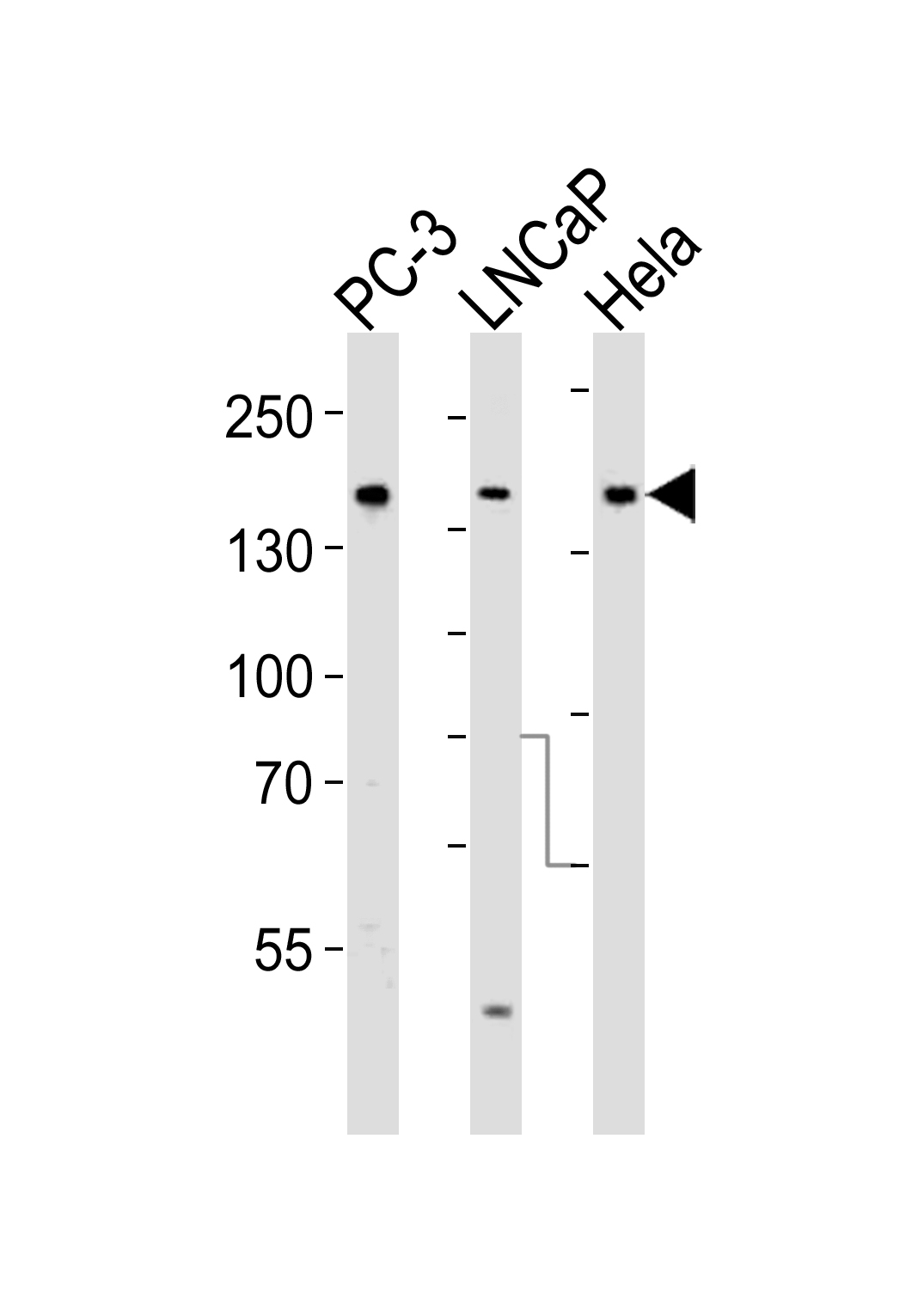
Western blot analysis of lysates from PC-3, LNCaP, Hela cell line (from left to right), using Tuberin (TSC2) Antibody(Cat. #JP110581). JP110581 was diluted at 1:1000 at each lane. A goat anti-rabbit IgG H&L(HRP) at 1:5000 dilution was used as the secondary antibody. Lysates at 35ug per lane.
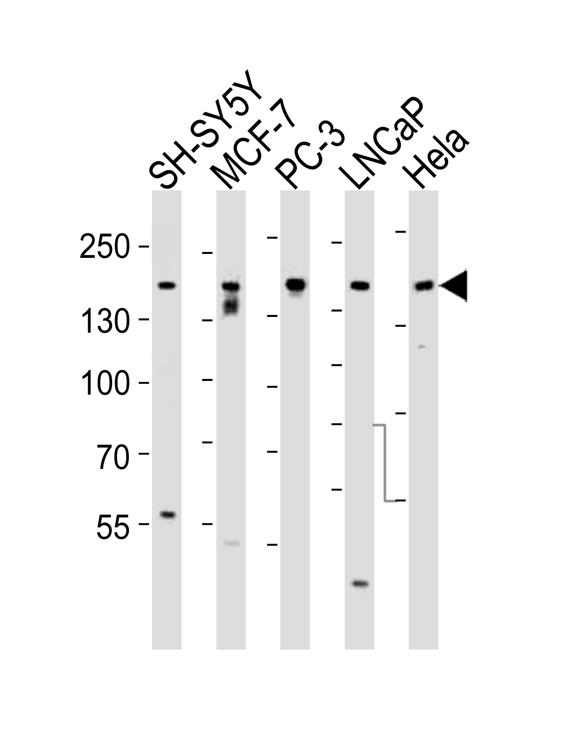
Western blot analysis of lysates from SH-SY5Y, MCF-7, PC-3, LNCaP, Hela, cell line (from left to right), using Tuberin (TSC2) Antibody(Cat. #JP110581). JP110581 was diluted at 1:1000 at each lane. A goat anti-rabbit IgG H&L(HRP) at 1:5000 dilution was used as the secondary antibody. Lysates at 35ug per lane.

Fluorescent confocal image of Hela cell stained with Tuberin (TSC2) Antibody (S1798)(Cat#JP110581).Hela cells were fixed with 4% PFA (20 min), permeabilized with Triton X-100 (0.1%, 10 min), then incubated with Tuberin (TSC2) primary antibody (1:25, 1 h at 37℃). For secondary antibody, Alexa Fluor® 488 conjugated donkey anti-rabbit antibody (green) was used (1:400, 50 min at 37℃).Cytoplasmic actin was counterstained with Alexa Fluor® 555 (red) conjugated Phalloidin (7units/ml, 1 h at 37℃). Nuclei were counterstained with DAPI (blue) (10 µg/ml, 10 min).Tuberin (TSC2) immunoreactivity is localized to Cytoplasm significantly.
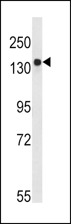
Western blot analysis of TSC2-pS1798 (Cat. #JP110581) in Ramos cell line lysates (35ug/lane). TSC2 (arrow) was detected using the purified Pab.

Formalin-fixed and paraffin-embedded human hepatocarcinoma tissue reacted with TSC2 Antibody (S1798) (Cat.#JP110581), which was peroxidase-conjugated to the secondary antibody, followed by DAB staining. This data demonstrates the use of this antibody for immunohistochemistry; clinical relevance has not been evaluated.