
Mouse anti-ERCC1 Monoclonal Antibody(C-term) (752CT13.2.5)描述别名宿主特异性反应种属应用分子量类型克隆号同种型储存/保存方法存储溶液研究领域背景说明细胞定位UniProt参考文献
| 概述 | |
| 描述 |
Mouse Monoclonal Antibody (Mab)
|
| 别名 |
ERCC1抗体;DNA excision repair protein ERCC-1; ERCC1
|
| 宿主 |
Mouse
|
| 特异性 |
This ERCC1 antibody is generated from mice immunized with a KLH conjugated synthetic peptide between 268-297 amino acids from the C-terminal region of human ERCC1.
|
| 反应种属 |
Human
|
| 应用 |
WB~~1:1000
|
| 分子量 |
Predicted molecular weight: 33kD
Disclaimer note: The observed molecular weight of the protein may vary from the listed predicted molecular weight due to post translational modifications, post translation cleavages, relative charges, and other experimental factors. |
| 性能 | |
| 类型 |
Monoclonal Antibody
|
| 克隆号 |
752CT13.2.5
|
| 同种型 |
IgM,k
|
| 储存/保存方法 |
Maintain refrigerated at 2-8°C for up to 2 weeks. For long term storage store at -20°C in small aliquots to prevent freeze-thaw cycles.
|
| 存储溶液 |
Purified monoclonal antibody supplied in PBS with 0.09% (W/V) sodium azide. This antibody is prepared by Euglobin precipitation followed by dialysis against PBS.
|
| 研究领域 |
Cancer;Crown Antibodies
|
| 靶标 | |
| 背景说明 |
Isoform 1: Non-catalytic component of a structure- specific DNA repair endonuclease responsible for the 5′-incision during DNA repair. Responsible, in conjunction with SLX4, for the first step in the repair of interstrand cross-links (ICL). Participates in the processing of anaphase bridge-generating DNA structures, which consist in incompletely processed DNA lesions arising during S or G2 phase, and can result in cytokinesis failure. Also required for homology-directed repair (HDR) of DNA double-strand breaks, in conjunction with SLX4.
|
| 细胞定位 |
Isoform 1: Nucleus. Isoform 3: Nucleus.
|
| UniProt |
P07992
|
| 参考文献 | |
| 参考文献 |
van Duin M., et al. Cell 44:913-923(1986).
Hoeijmakers J.H.J., et al. Cold Spring Harb. Symp. Quant. Biol. 51:91-101(1986). Yu J.J., et al. Mutat. Res. 382:13-20(1997). Hisatomi H., et al. Submitted (AUG-2001) to the EMBL/GenBank/DDBJ databases. Kalnine N., et al. Submitted (MAY-2003) to the EMBL/GenBank/DDBJ databases. |
实验结果图
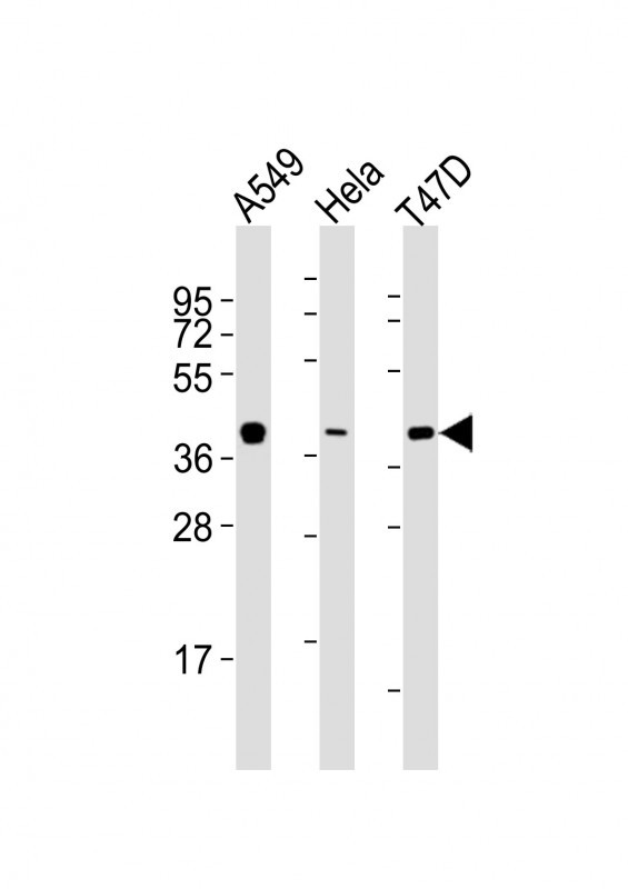
All lanes : Anti-ERCC1 Antibody (C-term) at 1:2000 dilution Lane 1: A549 whole cell lysates Lane 2: Hela whole cell lysates Lane 3: T47D whole cell lysates Lysates/proteins at 20 μg per lane. Secondary Goat Anti-mouse IgG, (H+L), Peroxidase conjugated at 1/10000 dilution Predicted band size : 33 kDa Blocking/Dilution buffer: 5% NFDM/TBST.
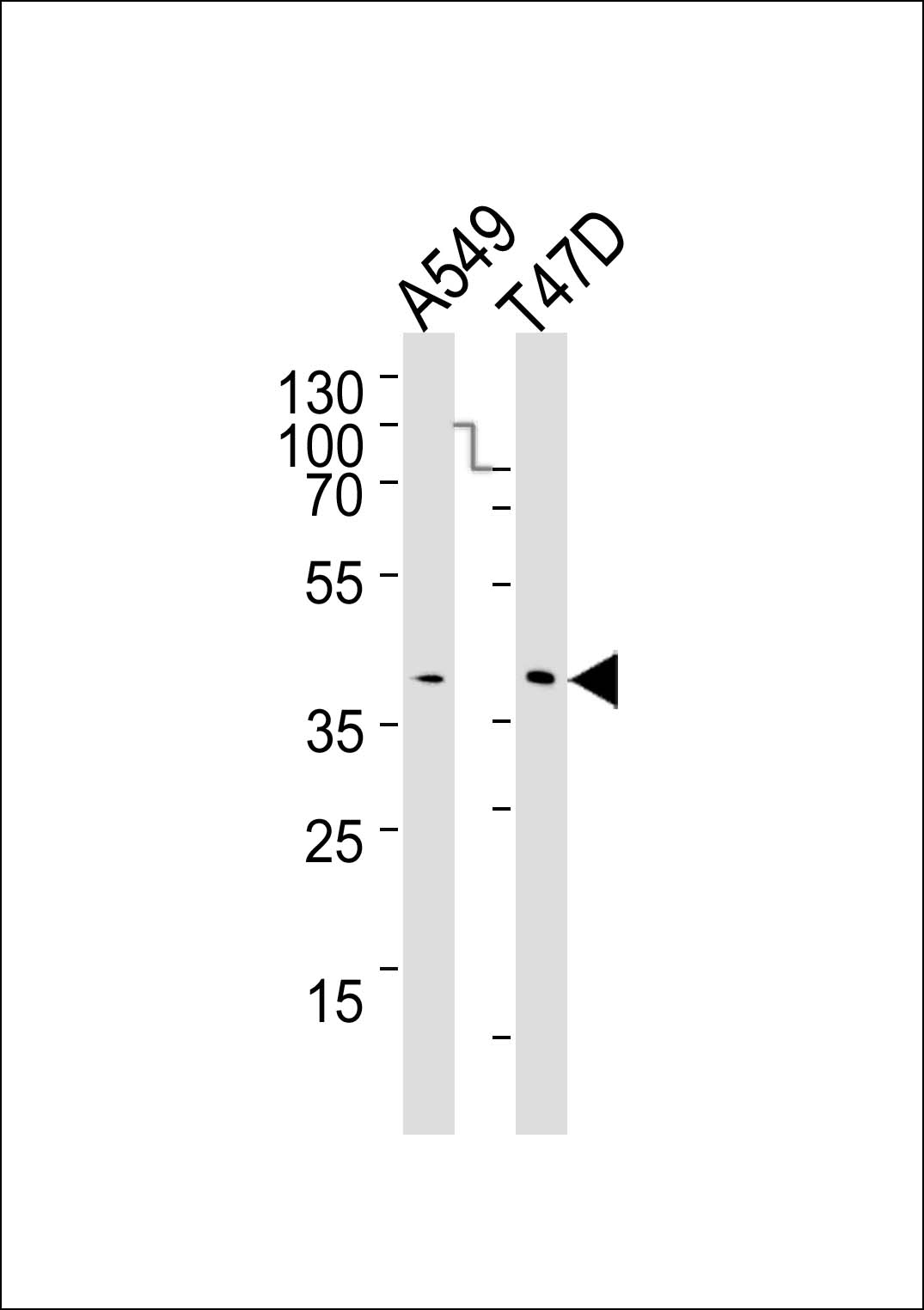
Western blot analysis of lysates from A549, T47D cell line (from left to right), using ERCC1 Antibody (C-term)(Cat. #JP100326). JP100326 was diluted at 1:1000 at each lane. A goat anti-mouse IgG H&L(HRP) at 1:3000 dilution was used as the secondary antibody. Lysates at 20μg per lane.
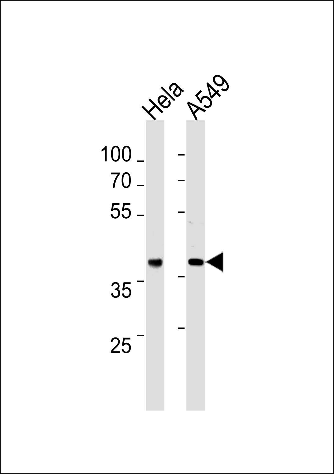
ERCC1 Antibody (C-term) (Cat. #JP100326) western blot analysis in Hela,A549 cell line lysates (35μg/lane).This demonstrates the ERCC1 antibody detected the ERCC1 protein (arrow).