
Mouse anti-EGFR Monoclonal Antibody(C-term) (688CT33.1.3)描述别名宿主特异性反应种属应用分子量类型克隆号同种型储存/保存方法存储溶液研究领域背景说明细胞定位UniProt参考文献
| 概述 | |
| 描述 |
Mouse Monoclonal Antibody (Mab)
|
| 别名 |
EGFR抗体;Epidermal growth factor receptor; Proto-oncogene c-ErbB-1; Receptor tyrosine-protein kinase erbB-1; EGFR; ERBB; ERBB1; HER1
|
| 宿主 |
Mouse
|
| 特异性 |
This EGFR antibody is generated from mice immunized with a KLH conjugated synthetic peptide between 1163-1191 amino acids from the C-terminal region of human EGFR.
|
| 反应种属 |
Human
|
| 应用 |
WB~~1:1000
IHC-P~~1:25 |
| 分子量 |
Predicted molecular weight: 134kD
Disclaimer note: The observed molecular weight of the protein may vary from the listed predicted molecular weight due to post translational modifications, post translation cleavages, relative charges, and other experimental factors. |
| 性能 | |
| 类型 |
Monoclonal Antibody
|
| 克隆号 |
688CT33.1.3
|
| 同种型 |
IgG1
|
| 储存/保存方法 |
Maintain refrigerated at 2-8°C for up to 2 weeks. For long term storage store at -20°C in small aliquots to prevent freeze-thaw cycles.
|
| 存储溶液 |
Purified monoclonal antibody supplied in PBS with 0.09% (W/V) sodium azide. This antibody is purified through a protein G column, eluted with high and low pH buffers and neutralized immediately, followed by dialysis against PBS.
|
| 研究领域 |
Cancer;Signal Transduction
|
| 靶标 | |
| 背景说明 |
Receptor tyrosine kinase binding ligands of the EGF family and activating several signaling cascades to convert extracellular cues into appropriate cellular responses. Known ligands include EGF, TGFA/TGF-alpha, amphiregulin, epigen/EPGN, BTC/betacellulin, epiregulin/EREG and HBEGF/heparin-binding EGF. Ligand binding triggers receptor homo- and/or heterodimerization and autophosphorylation on key cytoplasmic residues. The phosphorylated receptor recruits adapter proteins like GRB2 which in turn activates complex downstream signaling cascades. Activates at least 4 major downstream signaling cascades including the RAS- RAF-MEK-ERK, PI3 kinase-AKT, PLCgamma-PKC and STATs modules. May also activate the NF-kappa-B signaling cascade. Also directly phosphorylates other proteins like RGS16, activating its GTPase activity and probably coupling the EGF receptor signaling to the G protein-coupled receptor signaling. Also phosphorylates MUC1 and increases its interaction with SRC and CTNNB1/beta-catenin.
|
| 细胞定位 |
Cell membrane; Single-pass type I membrane protein. Endoplasmic reticulum membrane; Single-pass type I membrane protein. Golgi apparatus membrane; Single-pass type I membrane protein. Nucleus membrane; Single-pass type I membrane protein. Endosome. Endosome membrane. Nucleus. Note=In response to EGF, translocated from the cell membrane to the nucleus via Golgi and ER. Endocytosed upon activation by ligand. Colocalized with GPER1 in the nucleus of estrogen agonist-induced cancer-associated fibroblasts (CAF)
|
| UniProt |
P00533
|
| 参考文献 | |
| 参考文献 |
Abdallah, R.T., et al. J. Biol. Chem. 285(45):35206-35215(2010)
Lu, C., et al. Mol. Cell. Biol. 30(22):5432-5443(2010) Rosell, R., et al. Ann. N. Y. Acad. Sci. 1210, 45-52 (2010) : Hata, A., et al. J Thorac Oncol 5(10):1524-1528(2010) Aguirre, A., et al. Nature 467(7313):323-327(2010) |
实验结果图
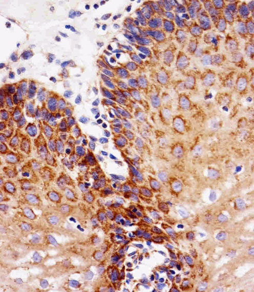
Immunohistochemical analysis of paraffin-embedded H. esophagus section using EGFR Antibody (C-term)(Cat#JP100322). JP100322 was diluted at 1:25 dilution. A peroxidase-conjugated goat anti-mouse IgG at 1:400 dilution was used as the secondary antibody, followed by DAB staining.
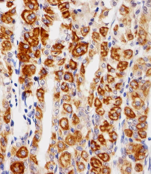
Immunohistochemical analysis of paraffin-embedded H. stomach section using EGFR Antibody (C-term)(Cat#JP100322). JP100322 was diluted at 1:25 dilution. A peroxidase-conjugated goat anti-mouse IgG at 1:400 dilution was used as the secondary antibody, followed by DAB staining.
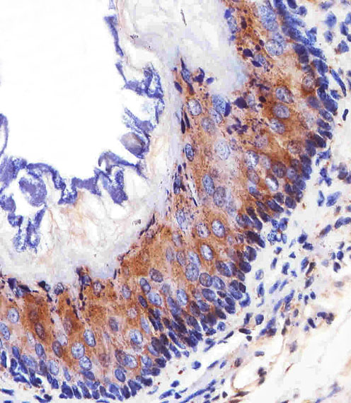
Immunohistochemical analysis of paraffin-embedded M. esophagus section using EGFR Antibody (C-term)(Cat#JP100322). JP100322 was diluted at 1:25 dilution. A peroxidase-conjugated goat anti-mouse IgG at 1:400 dilution was used as the secondary antibody, followed by DAB staining.
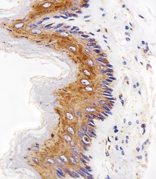
Immunohistochemical analysis of paraffin-embedded R. esophagus section using EGFR Antibody (C-term)(Cat#JP100322). JP100322 was diluted at 1:25 dilution. A peroxidase-conjugated goat anti-mouse IgG at 1:400 dilution was used as the secondary antibody, followed by DAB staining.
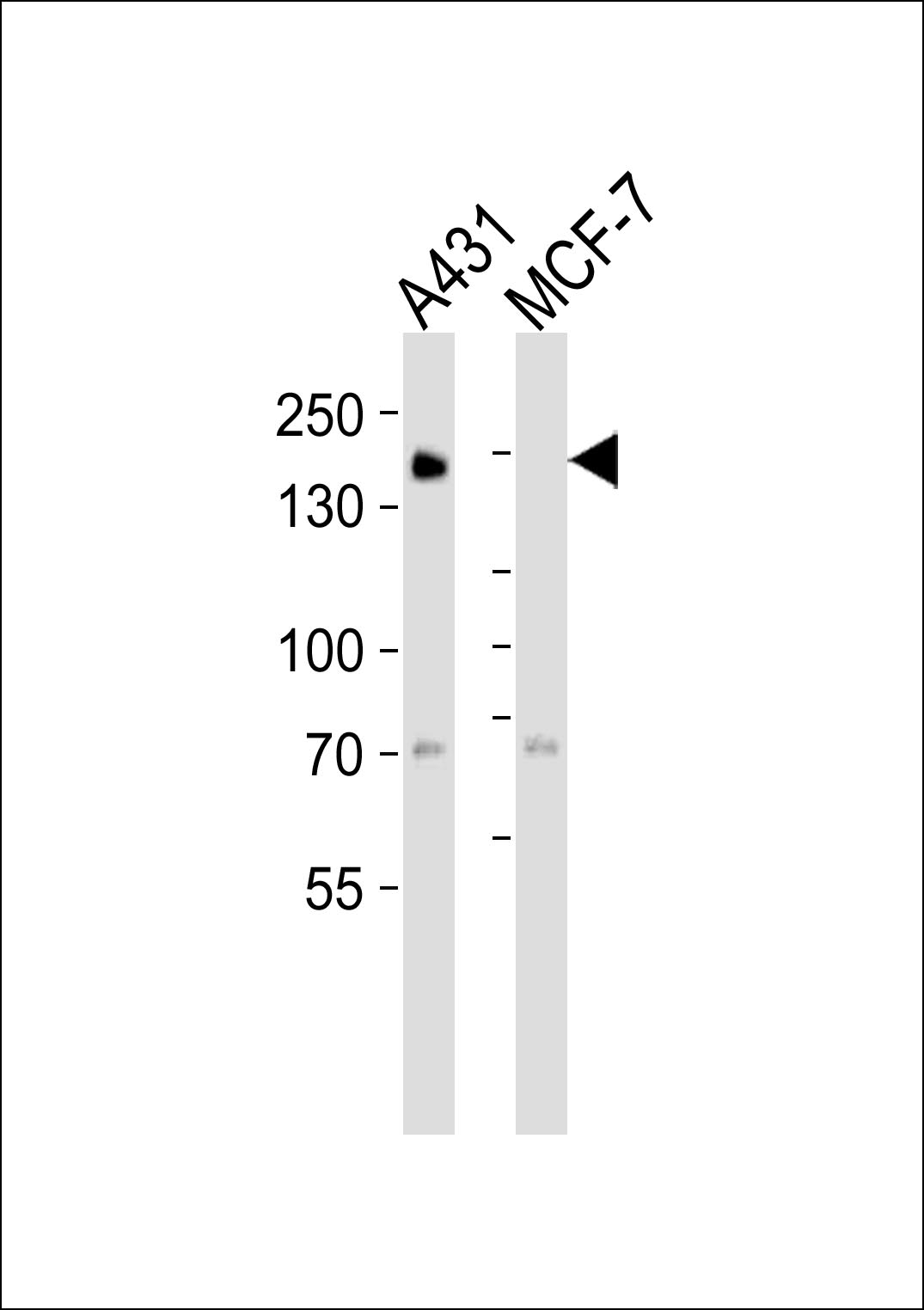
Western blot analysis of lysates from A431, MCF-7 cell line (from left to right), using EGFR Antibody (C-term) (Cat. #JP100322). JP100322 was diluted at 1:1000 at each lane. A goat anti-mouse IgG H&L(HRP) at 1:3000 dilution was used as the secondary antibody. Lysates at 35μg per lane.
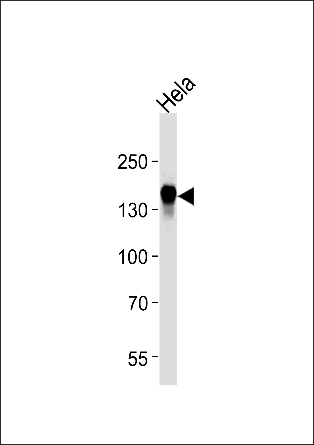
EGFR Antibody (C-term) (Cat. #JP100322) western blot analysis in Hela cell line lysates (35μg/lane).This demonstrates the EGFR antibody detected the EGFR protein (arrow).