
Mouse anti-KIT Monoclonal Antibody(566CT8.5.4)描述别名宿主特异性反应种属应用分子量类型克隆号同种型储存/保存方法存储溶液研究领域背景说明细胞定位UniProt参考文献
| 概述 | |
| 描述 |
Mouse Monoclonal Antibody (Mab)
|
| 别名 |
KIT抗体;Mast/stem cell growth factor receptor Kit; SCFR; Piebald trait protein; PBT; Proto-oncogene c-Kit; Tyrosine-protein kinase Kit; p145 c-kit; v-kit Hardy-Zuckerman 4 feline sarcoma viral oncogene homolog; CD117; KIT
|
| 宿主 |
Mouse
|
| 特异性 |
This KIT Antibody is generated from mouses immunized with human KIT recombinant protein.
|
| 反应种属 |
Human
|
| 应用 |
IF~~1:10~50
WB~~1:100 |
| 分子量 |
Predicted molecular weight: 110kD
Disclaimer note: The observed molecular weight of the protein may vary from the listed predicted molecular weight due to post translational modifications, post translation cleavages, relative charges, and other experimental factors. |
| 性能 | |
| 类型 |
Monoclonal Antibody
|
| 克隆号 |
566CT8.5.4
|
| 同种型 |
IgM
|
| 储存/保存方法 |
Maintain refrigerated at 2-8°C for up to 2 weeks. For long term storage store at -20°C in small aliquots to prevent freeze-thaw cycles.
|
| 存储溶液 |
Purified monoclonal antibody supplied in PBS with 0.09% (W/V) sodium azide. This antibody is prepared by Euglobin precipitation followed by dialysis against PBS.
|
| 研究领域 |
Cancer;Developmental Biology;Immunology;Neuroscience;Signal Transduction
|
| 靶标 | |
| 背景说明 |
Tyrosine-protein kinase that acts as cell-surface receptor for the cytokine KITLG/SCF and plays an essential role in the regulation of cell survival and proliferation, hematopoiesis, stem cell maintenance, gametogenesis, mast cell development, migration and function, and in melanogenesis. In response to KITLG/SCF binding, KIT can activate several signaling pathways. Phosphorylates PIK3R1, PLCG1, SH2B2/APS and CBL. Activates the AKT1 signaling pathway by phosphorylation of PIK3R1, the regulatory subunit of phosphatidylinositol 3-kinase. Activated KIT also transmits signals via GRB2 and activation of RAS, RAF1 and the MAP kinases MAPK1/ERK2 and/or MAPK3/ERK1. Promotes activation of STAT family members STAT1, STAT3, STAT5A and STAT5B. Activation of PLCG1 leads to the production of the cellular signaling molecules diacylglycerol and inositol 1,4,5-trisphosphate. KIT signaling is modulated by protein phosphatases, and by rapid internalization and degradation of the receptor. Activated KIT promotes phosphorylation of the protein phosphatases PTPN6/SHP-1 and PTPRU, and of the transcription factors STAT1, STAT3, STAT5A and STAT5B. Promotes phosphorylation of PIK3R1, CBL, CRK (isoform Crk-II), LYN, MAPK1/ERK2 and/or MAPK3/ERK1, PLCG1, SRC and SHC1.
|
| 细胞定位 |
Isoform 1: Cell membrane; Single-pass type I membrane protein Isoform 3: Cytoplasm. Note=Detected in the cytoplasm of spermatozoa, especially in the equatorial and subacrosomal region of the sperm head
|
| UniProt |
P10721
|
| 参考文献 | |
| 参考文献 |
Molderings, G.J., et al. Immunogenetics 62 (11-12), 721-727 (2010) :
Cheng, M., et al. Circ. Res. 107(9):1083-1093(2010) Chi, P., et al. Nature 467(7317):849-853(2010) Rossi, S., et al. Am. J. Surg. Pathol. 34(10):1480-1491(2010) Chen, P., et al. World J. Gastroenterol. 16(33):4227-4232(2010) |
实验结果图
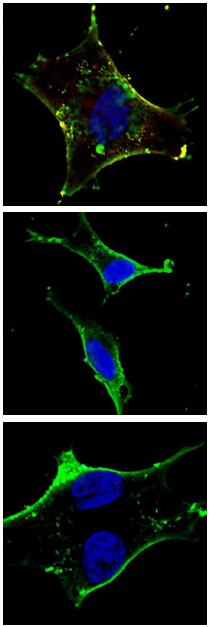
Immunofluorescent analysis of U251 cells, using KIT Antibody (Cat. #JP100295). JP100295 was diluted at 1:100 dilution. Dylight Fluor 488-conjugated goat anti-mouse lgG at 1:400 dilution was used as the secondary antibody (green). Cytoplasmic actin was counterstained with Dylight Fluor® 554 (red) conjugated Phalloidin (red).

KIT Antibody(Cat. #JP100295) western blot analysis in A549 cell line lysates (35μg/lane).This demonstrates the KIT antibody detected the KIT protein (arrow).
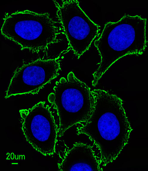
Fluorescent confocal image of an SY5Y (SH-SY5Y; ATCC#CRL-2266) cell stained with JP100295 KIT antibody. SY5Y cells were fixed with 4% PFA (20 min), permeabilized with Triton X-100 (0.2%, 30 min), then incubated with JP100295 KIT primary antibody (1:500, 2 h at room temperature). For secondary antibody, Alexa Fluor® 488 conjugated donkey anti-mouse antibody (green) was used (1:1000, 1h). Cytoplasmic actin was counterstained with Alexa Fluor® 555 (red) conjugated Phalloidin (5.25 μM, 25 min). Nuclei were counterstained with Hoechst 33342 (blue) (10 μg/ml, 3 min). Note the highly specific localization of the KIT immunosignal to the membranes, especially the plasma membrane. ibid. References for SY5Y (SH-SY5Y; ATCC#CRL-2266): 1. Ross RA, et al. Coordinate morphological and biochemical interconversion of human neuroblastoma cells. J. Natl. Cancer Inst. 71: 741-749, 1983. [PubMed: 6137586]; 2. Biedler JL, et al. Multiple neurotransmitter synthesis by human neuroblastoma cell lines and clones. Cancer Res. 38: 3751-3757, 1978. [PubMed: 29704]. Ibid.