
Rabbit anti-p38 MAPK Recombinant Monoclonal Antibody(S-508-19)描述别名宿主特异性反应种属预测反应种属应用免疫原形式浓度纯化方法类型克隆号储存/保存方法存储溶液背景说明组织特异性翻译后修饰细胞定位UniProt
| 概述 | |
| 描述 |
The protein encoded by this gene is a member of the MAP kinase family. MAP kinases act as an integration point for multiple biochemical signals, and are involved in a wide variety of cellular processes such as proliferation, differentiation, transcription regulation and development.
|
| 别名 |
p38 MAPK抗体;Mitogen-activated protein kinase 14; MAP kinase 14; MAPK 14; CSAID-binding protein; CSBP; MAP kinase MXI2; MAX-interacting protein 2; MAP kinase p38 alpha; SAPK2a
|
| 宿主 |
Rabbit
|
| 特异性 |
p38 MAPK Antibody detects endogenous levels of total p38 MAPK.
|
| 反应种属 |
Human, Mouse, Rat
|
| 预测反应种属 |
Pig;Rabbit;Sheep;Dog;Horse;Bovine;
|
| 应用 |
WB: 1:5000, IHC-P: 1:1000, ICC: 1:500, FC(Intra): 1:500
|
| 免疫原 |
Synthetic peptide
|
| 性能 | |
| 形式 |
Liquid
|
| 浓度 |
0.5 mg/mL
|
| 纯化方法 |
Protein A affinity column
|
| 类型 |
Monoclonal Antibody
|
| 克隆号 |
S-508-19
|
| 储存/保存方法 |
Store at -20℃ for one year.
|
| 存储溶液 |
PBS, 40% Glycerol, 0.05% BSA, 0.03% Proclin 300
|
| 靶标 | |
| 背景说明 |
p38 mitogen-activated protein kinases are a class of mitogen-activated protein kinases (MAPKs) that are responsive to stress stimuli, such as cytokines, ultraviolet irradiation, heat shock, and osmotic shock, and are involved in cell differentiation, apoptosis and autophagy. Abnormal activity (higher or lower than physiological) of p38 has been implicated in pathological stresses in several tissues, that include neuronal, bone, lung, cardiac and skeletal muscle, red blood cells, and fetal tissues.
|
| 组织特异性 |
Brain, heart, placenta, pancreas and skeletal muscle. Expressed to a lesser extent in lung, liver and kidney.
|
| 翻译后修饰 |
Dually phosphorylated on Thr-180 and Tyr-182 by the MAP2Ks MAP2K3/MKK3, MAP2K4/MKK4 and MAP2K6/MKK6 in response to inflammatory citokines, environmental stress or growth factors, which activates the enzyme. Dual phosphorylation can also be mediated by TAB1-mediated autophosphorylation. TCR engagement in T-cells also leads to Tyr-323 phosphorylation by ZAP70. Dephosphorylated and inactivated by DUPS1, DUSP10 and DUSP16. PPM1D also mediates dephosphorylation and inactivation of MAPK14 (PubMed:21283629).Acetylated at Lys-53 and Lys-152 by KAT2B and EP300. Acetylation at Lys-53 increases the affinity for ATP and enhances kinase activity. Lys-53 and Lys-152 are deacetylated by HDAC3.Ubiquitinated. Ubiquitination leads to degradation by the proteasome pathway.
|
| 细胞定位 |
Cytoplasm, Nucleus
|
| UniProt |
Q16539
|
实验结果图
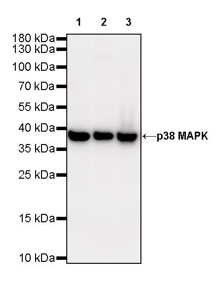
WB result of p38 MAPK Rabbit mAb Primary antibody: p38 MAPK Rabbit mAb at 1/5000 dilution Lane 1: HeLa whole cell lysate 20 ug Lane 2: Jurkat whole cell lysate 20 ug Lane 3: MCF7 whole cell lysate 20 ug Secondary antibody: Goat Anti-Rabbit IgG, (H+L), HRP conjugated at 1/10000 dilution Predicted MW: 41 kDa Observed MW: 38 kDa Exposure time: 90 s

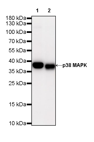
WB result of p38 MAPK Rabbit mAb Primary antibody: p38 MAPK Rabbit mAb at 1/5000 dilution Lane 1: NIH/3T3 whole cell lysate 20 ug Lane 2: mouse spleen lysate 20 ug Secondary antibody: Goat Anti-Rabbit IgG, (H+L), HRP conjugated at 1/10000 dilution Predicted MW: 41 kDa Observed MW: 38 kDa Exposure time: 180 s
WB result of p38 MAPK Rabbit mAb Primary antibody: p38 MAPK Rabbit mAb at 1/5000 dilution Lane 1: C6 whole cell lysate 20 ug Lane 2: rat spleen lysate 20 ug Secondary antibody: Goat Anti-Rabbit IgG, (H+L), HRP conjugated at 1/10000 dilution Predicted MW: 41 kDa Observed MW: 38 kDa Exposure time: 180 s
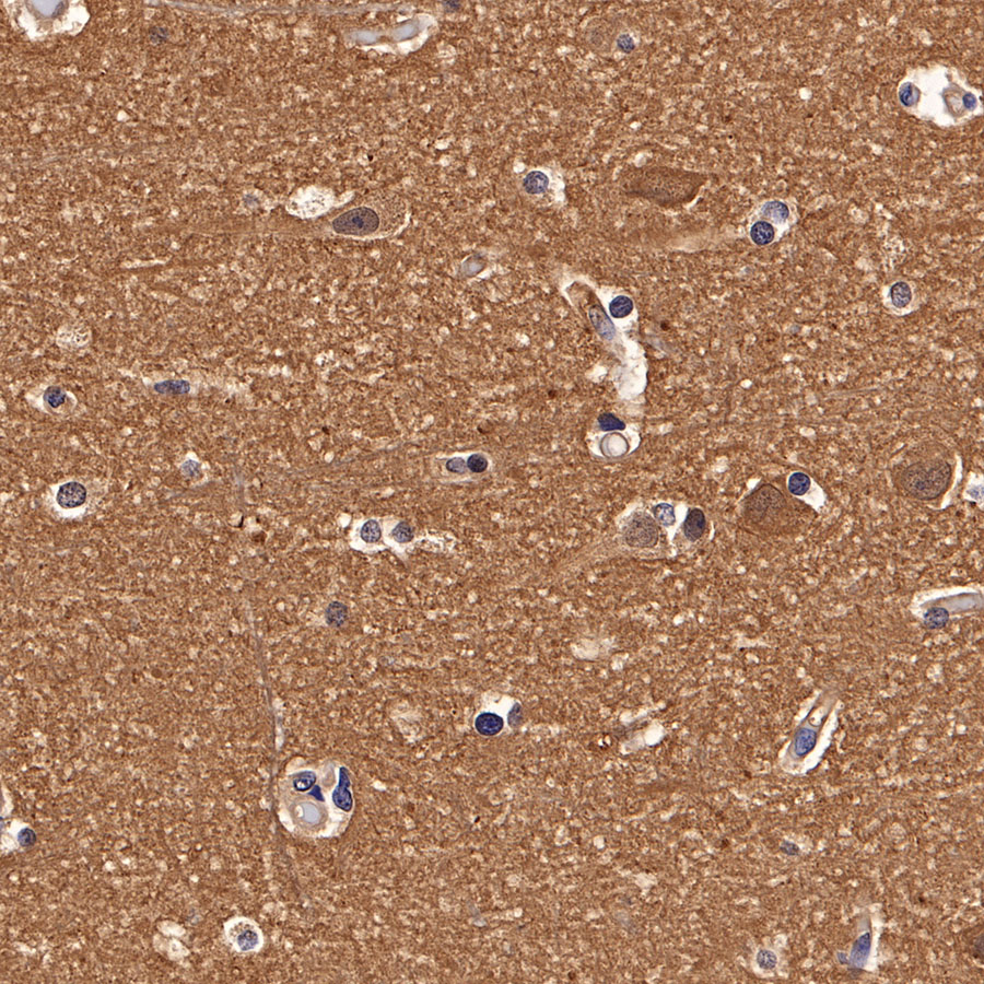
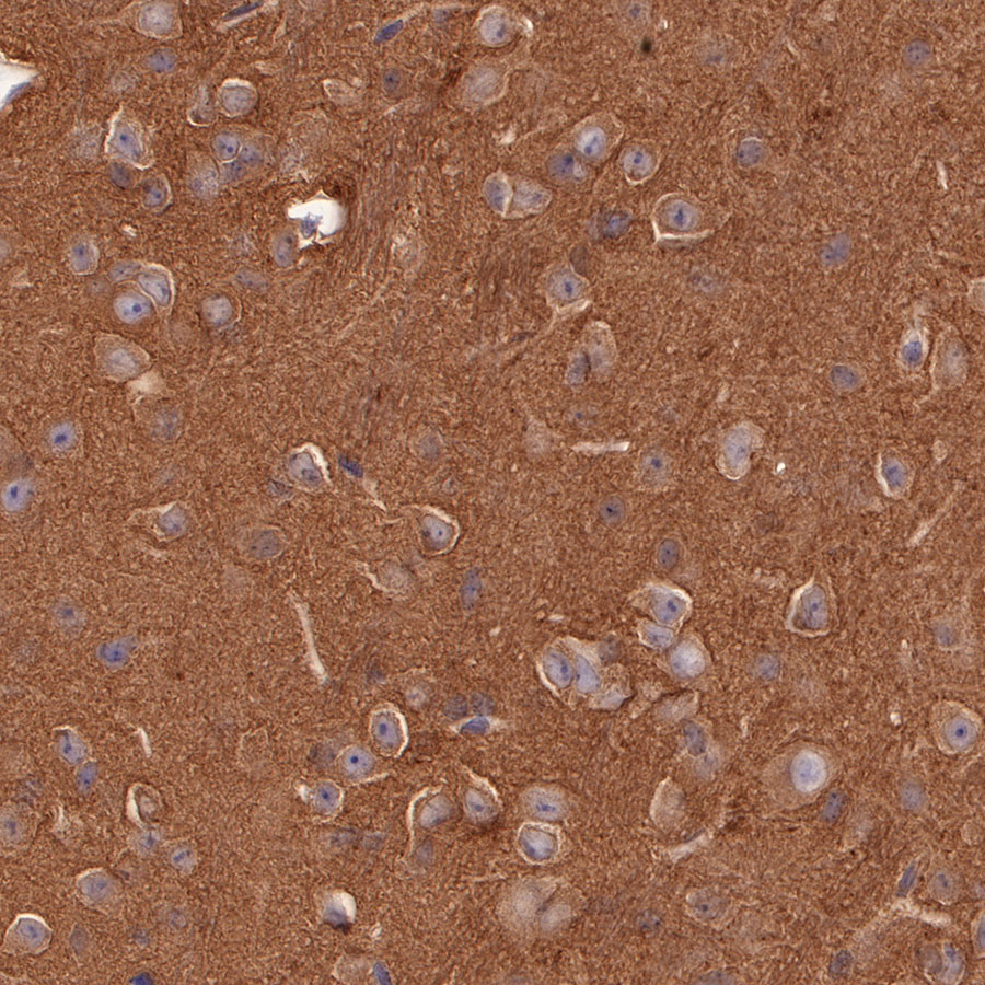
IHC shows positive staining in paraffin-embedded human cerebral cortex. Anti-p38 MAPK antibody was used at 1/1000 dilution, followed by a HRP Polymer for Mouse & Rabbit IgG (ready to use). Counterstained with hematoxylin. Heat mediated antigen retrieval with Tris/EDTA buffer pH9.0 was performed before commencing with IHC staining protocol.
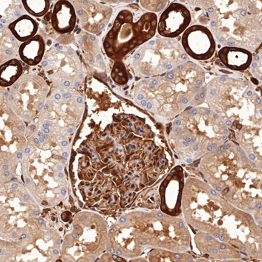
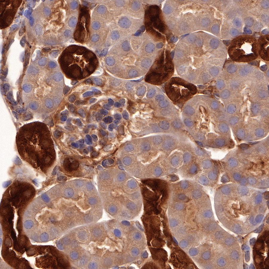
IHC shows positive staining in paraffin-embedded human kidney. Anti-p38 MAPK antibody was used at 1/1000 dilution, followed by a HRP Polymer for Mouse & Rabbit IgG (ready to use). Counterstained with hematoxylin. Heat mediated antigen retrieval with Tris/EDTA buffer pH9.0 was performed before commencing with IHC staining protocol.
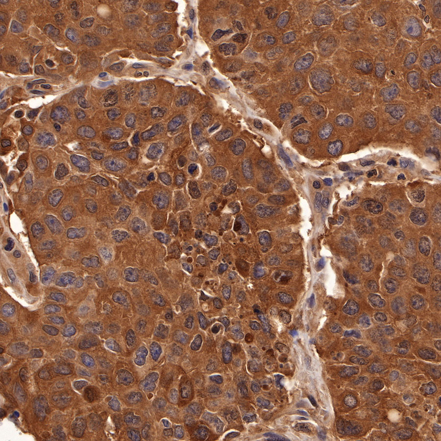
IHC shows positive staining in paraffin-embedded human breast cancer. Anti-p38 MAPK antibody was used at 1/1000 dilution, followed by a HRP Polymer for Mouse & Rabbit IgG (ready to use). Counterstained with hematoxylin. Heat mediated antigen retrieval with Tris/EDTA buffer pH9.0 was performed before commencing with IHC staining protocol.
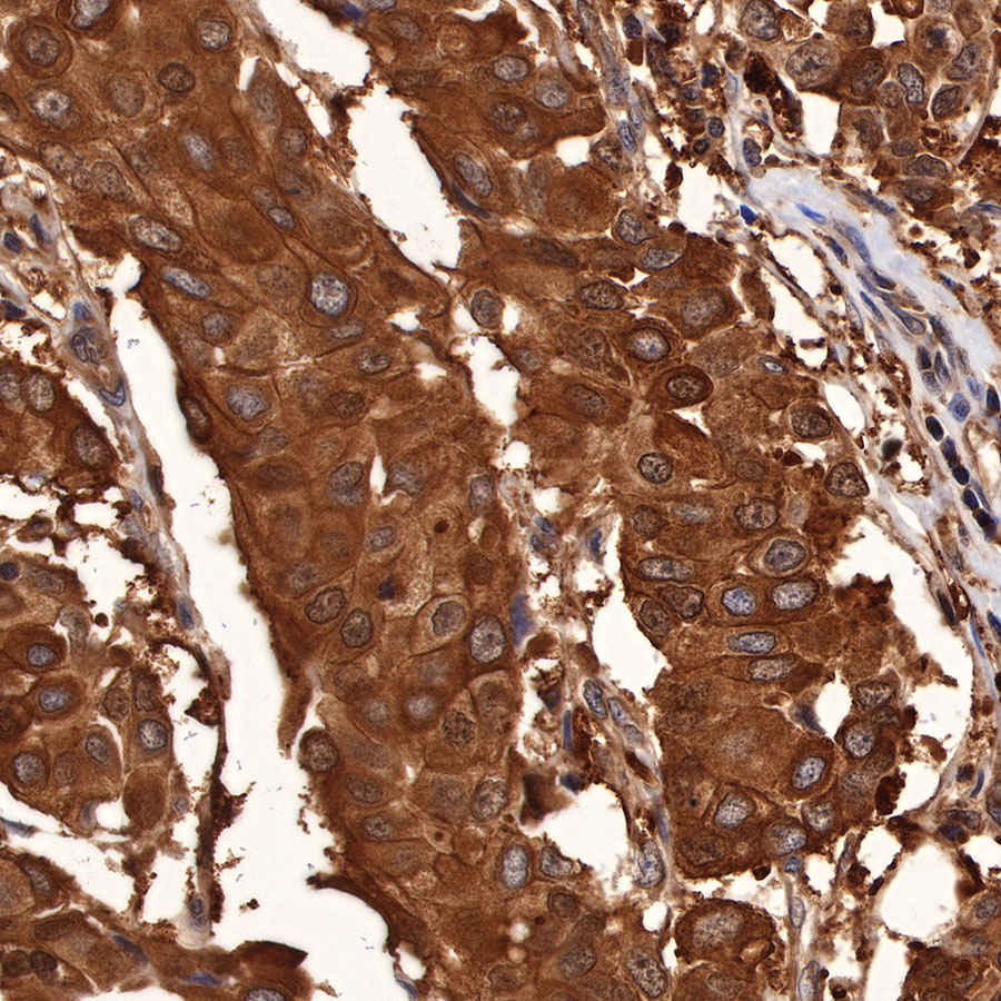
IHC shows positive staining in paraffin-embedded human lung squamous cell carcinoma. Anti-p38 MAPK antibody was used at 1/1000 dilution, followed by a HRP Polymer for Mouse & Rabbit IgG (ready to use). Counterstained with hematoxylin. Heat mediated antigen retrieval with Tris/EDTA buffer pH9.0 was performed before commencing with IHC staining protocol.
IHC shows positive staining in paraffin-embedded mouse cerebral cortex. Anti-p38 MAPK antibody was used at 1/1000 dilution, followed by a HRP Polymer for Mouse & Rabbit IgG (ready to use). Counterstained with hematoxylin. Heat mediated antigen retrieval with Tris/EDTA buffer pH9.0 was performed before commencing with IHC staining protocol.
IHC shows positive staining in paraffin-embedded mouse kidney. Anti-p38 MAPK antibody was used at 1/1000 dilution, followed by a HRP Polymer for Mouse & Rabbit IgG (ready to use). Counterstained with hematoxylin. Heat mediated antigen retrieval with Tris/EDTA buffer pH9.0 was performed before commencing with IHC staining protocol.

IHC shows positive staining in paraffin-embedded rat cerebral cortex. Anti-p38 MAPK antibody was used at 1/1000 dilution, followed by a HRP Polymer for Mouse & Rabbit IgG (ready to use). Counterstained with hematoxylin. Heat mediated antigen retrieval with Tris/EDTA buffer pH9.0 was performed before commencing with IHC staining protocol.
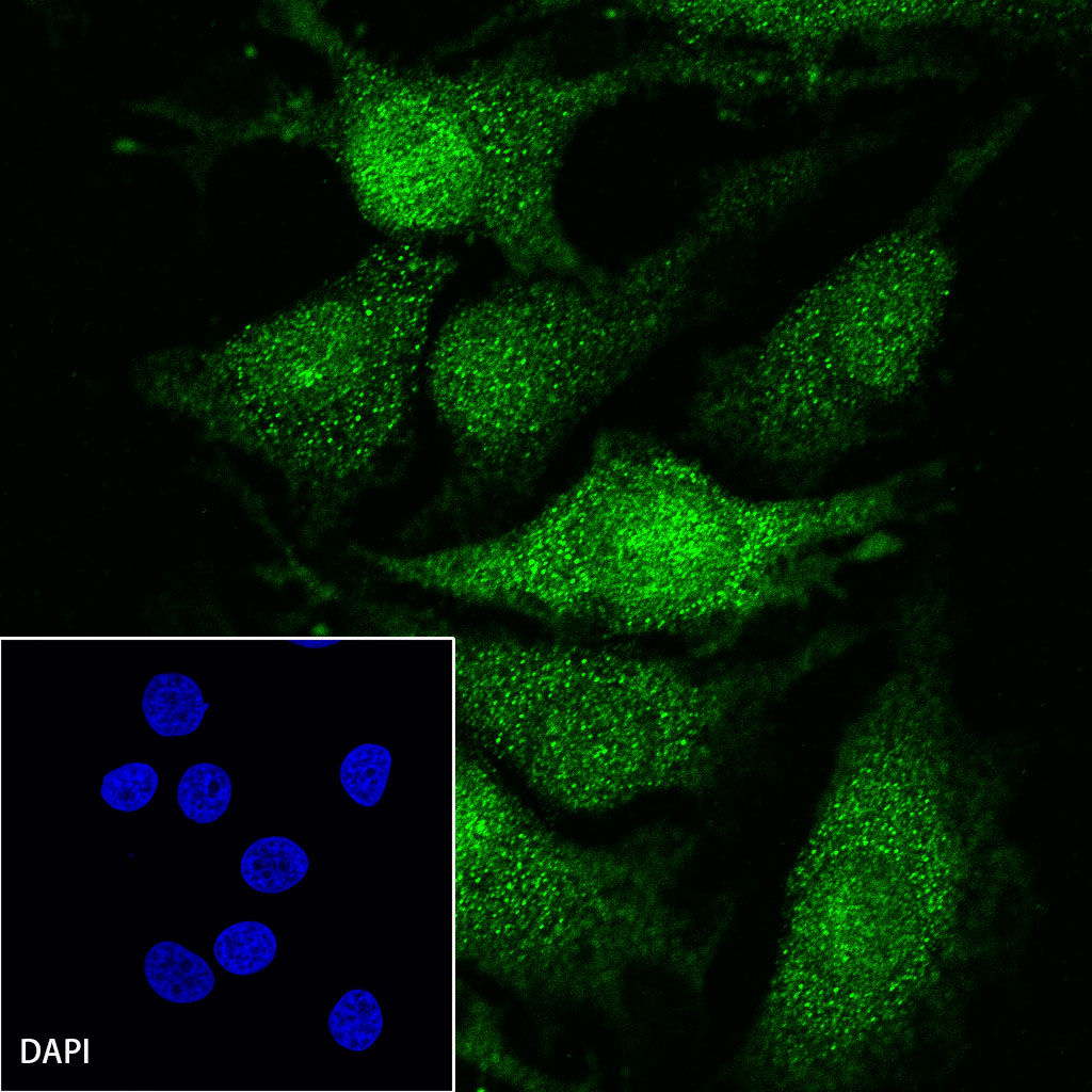
ICC shows positive staining in HeLa cells. Anti-p38 MAPK antibody was used at 1/500 dilution (Green) and incubated overnight at 4°C. Goat polyclonal Antibody to Rabbit IgG – H&L (Alexa Fluor® 488) was used as secondary antibody at 1/1000 dilution. The cells were fixed with 4% PFA and permeabilized with 0.1% PBS-Triton X-100. Nuclei were counterstained with DAPI.
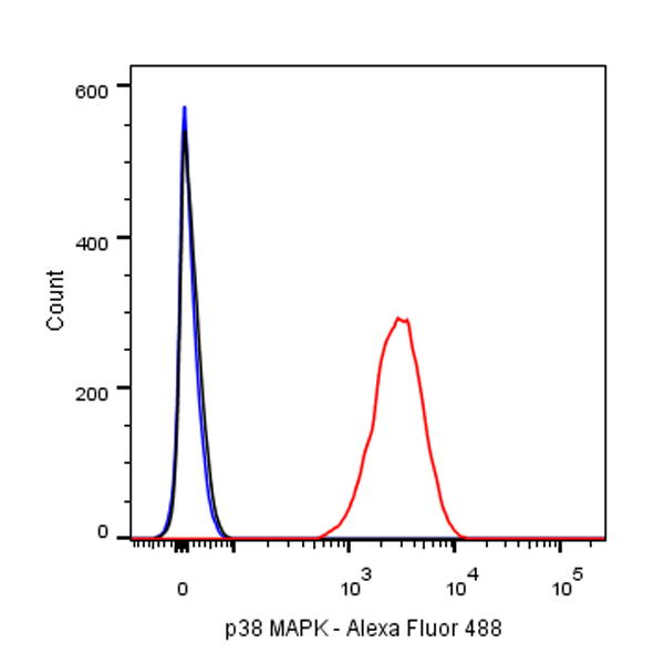
Flow cytometric analysis of 4% PFA fixed 90% methanol permeabilized HeLa (Human cervix adenocarcinoma epithelial cell) cells labelling p38 MAPK antibody at 1/500 dilution (0.1 μg) / (red) compared with a Rabbit monoclonal IgG isotype control (Black) and an unlabelled control (cells without incubation with primary antibody and secondary antibody) (Blue). Goat Anti – Rabbit IgG Alexa Fluor 488 was used as the secondary antibody.