
Bring fluorescence microscopy out of the darkroom and into your classroom. ZOE’s intuitive touch-screen interface, integrated digital camera, and projector connection port allow every student to experience fluorescence cell imag
描述
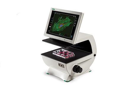
Take cell imaging out of the darkroom and into your classroom! The ZOE Fluorescent Cell Imager eliminates the complexities associated with traditional cell imaging microscopy. With the ZOE Cell Imager, you no longer have to squeeze all your students into a tiny darkroom, worry about damage to expensive shared research equipment, and waste precious time while each individual student takes a turn looking through a microscope lens.
The ZOE Fluorescent Cell Imager is a complete digital imaging system, allowing students to view samples, capture and store images, and create multicolor overlays. Thanks to the built-in light shield, the ZOE Cell Imager does not require a darkroom for fluorescence imaging.
The ZOE Cell Imager uses an intuitive touch-screen interface to control the brightfield, three fluorescence channels, and the integrated digital camera. Any student that has used a personal tablet or smartphone will be able to easily navigate the ZOE Imager’s controls.
The ZOE Imager’s 10.1″ (26 cm) screen and HDMI projector connectivity allows all your students to see the same live image at the same time. With a compact 13″ x 12.6″ (33 x 32 cm) footprint, you can easily transport the ZOE Imager from a research lab to a teaching lab or a classroom.
Take the ZOE Imager for a Test Drive
Both you and your students can take ZOE for a test drive. Just launch the ZOE Interactive Web App to try the intuitive ZOE user interface, manipulate images, view features and specifications in depth, and more.
Features and Benefits of the ZOE Fluorescent Cell Imager
- Fluorescence imaging in your classroom — light shield permits fluorescence imaging in ambient light and eliminates the need for a darkroom
- Simplified cell imaging — the intuitive touch-screen interface allows users to view cells, capture images, and create multichannel merges with minimal training
- HDMI connection — project live microscopy images, allowing the entire class (including online students) to see the same thing at the same time
- Flexible operation — brightfield and three fluorescence channels enable your students to learn both simple and sophisticated imaging techniques
- Robust construction — fully integrated system with long-life LEDs, ready for intensive daily use
- Small footprint — compact size accommodates crowded lab benches
Learn more about the features of the ZOE Cell Imager.
Applications of the ZOE Fluorescent Cell Imager
With ZOE’s brightfield and three fluorescent channels, your students can experience cell culture work as well as fluorescent techniques practiced regularly in the research lab:
- Observation of general cell health and morphology
- Monitoring of cell growth and proliferation
- Estimation of transfection efficiency
- Visualization of expressed fluorescent proteins
- Immunofluorescent protein localization
- Visual estimation of cell confluency
- Capturing cell images (with or without fluorescent labels)
Ways to Use Fluorophores with the ZOE Cell Imager in Your Classroom
Here are a few ways you can use the ZOE Imager in your classroom and teaching labs. Be creative and share your teaching ideas with other educators just like you at the Explorer Community. This is not a comprehensive list; other dyes and fluorescent proteins with compatible excitation and emission spectra can also be used.
| ZOE-Compatible Fluorophores | |||
| Blue Channel Excitation: 355/40 nm Emission: 433/36 nm |
Green Channel Excitation: 480/17 nm Emission: 517/23 nm |
Red Channel Excitation: 556/20 nm Emission: 615/61 nm |
Teaching Applications |
| PureBlu™ DAPI Nuclear Staining Dye PureBlu Hoechst 33342 |
ER-Tracker Green MitoTracker Green FM |
ER-Tracker Red MitoTracker Red* |
These organelle-specific stains can help you teach cell structure to your students. Use several stains in combination with the ZOE Imager to create beautiful multicolored images that will inspire curiosity in your students. |
| VivaFix™ 353/442 Cell Viability Assay |
VivaFix 498/521 Cell Viability Assay* |
VivaFix 547/573 Cell Viability Assay VivaFix 583/603 |
These assays will allow your students to test the effects of environmental conditions, drugs, and chemicals on cell viability. |
| CFDA-SE | Your students can follow cell proliferation using this dye. They can visualize up to 8 cell divisions if they have the patience for it. | ||
| ReadiLink 350/440 Antibody Labeling Kit |
ReadiLink 492/516 Antibody Labeling Kit |
ReadiLink 555/570 Antibody Labeling Kit ReadiLink 594/610 |
Already have primary antibodies from your research lab? Share your research with your students using these fluorescent secondary antibody labeling kits. |
| mKate* RFP* |
EGFP* | mCherry* mStrawberry YFP* |
Use these fluorescent proteins to help your students learn about gene expression, tissue-specific protein expression, developmental biology, and genetics; compare mutant with wild-type organisms; and much more. |
| Alexa Fluor 488 dye* phalloidin | Tubulin Green | Use these tubulin- and actin-specific dyes on various species to help you teach comparative morphology in the classroom and lab. | |
| Other ZOE-Compatible Fluorophores | |||
| Alexa Fluor 350 | Acridine Orange | Alexa Fluor 546 | |
| Alexa Fluor 405 | BODIPY Fl* | Alexa Fluor 568 | |
| Cascade Blue | Calcein AM | Alexa Fluor 594 | |
| CellTracker Blue | DiO | Alexa Fluor 610 | |
| DAPI* | FITC* | Cy3* | |
| Hoechst* | SYTO 9, SYTO 13, SYTO 16 | Dil Stain | |
| Marina Blue | SYTOX Green | DsRed* | |
| SYTOX Orange | |||
| SYTO 84, SYTO 85 | |||
| Texas Red* | |||
* R&D Tested
技术指标
Green channel: blue LED
Red channel: green LED
Brightfield channel: multiple green LEDs (reduces chromatic aberration)
1,280 x 768 pixel image resolution, 80–180° angle tilt range
Multiwell plates: 6-, 12-, 24-, 48-, 96-, or 384-well microplates
Dishes: 35 mm, 60 mm, or 100 mm
Slides: chamber slides or standard glass microscopy slides
试剂&试剂盒
1665111EDU

1665112EDU
相关资料
Login Required
ZOE™ Fluorescent Cell Imager Brochure, Rev A
[ Add to Cart (Free) ]
ZOE Fluorescent Cell Imager Instruction Manual
ZOE™ Fluorescent Cell Imager Quick Guide, Rev B
ZOE: The Perfect Teaching Assistant Flier, Rev A
Fluorescent Cell Imaging Activities for Your Classroom Flier, Ver B