
Rabbit anti-Synaptophysin Recombiant Monoclonal Antibody(137-21)描述别名宿主特异性反应种属预测反应种属应用免疫原形式浓度纯化方法类型克隆号储存/保存方法存储溶液背景说明组织特异性翻译后修饰细胞定位UniProt
| 概述 | |
| 描述 |
SYP Possibly involved in structural functions as organizing other membrane components or in targeting the vesicles to the plasma membrane. Involved in the regulation of short-term and long-term synaptic plasticity. Belongs to the synaptophysin/synaptobrevin family. Homohexamer or homotetramer. Note: This description may include information from UniProtKB.
|
| 别名 |
Synaptophysin抗体;Major synaptic vesicle protein p38; SYP
|
| 宿主 |
Rabbit
|
| 特异性 |
Synaptophysin antibody detects endogenous levels of total Synaptophysin.
|
| 反应种属 |
Human, Mouse, Rat
|
| 预测反应种属 |
Pig;Sheep;Bovine;Horse;
|
| 应用 |
WB: 1:500, IHC:1:1000(Hu), IHC: 1:4000(Ms,Rat), FC(Intra):1:500
|
| 免疫原 |
Synthetic peptide
|
| 性能 | |
| 形式 |
liquid
|
| 浓度 |
0.25mg/ml
|
| 纯化方法 |
Protein A affinity column
|
| 类型 |
Monoclonal antibody
|
| 克隆号 |
137-21
|
| 储存/保存方法 |
Store at -20℃ for one year.
|
| 存储溶液 |
PBS, 40% Glycerol, 0.05% BSA, 0.03% Proclin 300
|
| 靶标 | |
| 背景说明 |
Synaptophysin, also known as the major synaptic vesicle protein p38, is a protein that in humans is encoded by the SYP gene. The protein is a synaptic vesicle glycoprotein with four transmembrane domains weighing 38kDa. It is present in neuroendocrine cells and in virtually all neurons in the brain and spinal cord that participate in synaptic transmission. It acts as a marker for neuroendocrine tumors, and its ubiquity at the synapse has led to the use of synaptophysin immunostaining for quantification of synapses.
|
| 组织特异性 |
Characteristic of a type of small (30-80 nm) neurosecretory vesicles, including presynaptic vesicles, but also vesicles of various neuroendocrine cells of both neuronal and epithelial phenotype.
|
| 翻译后修饰 |
Ubiquitinated; mediated by SIAH1 or SIAH2 and leading to its subsequent proteasomal degradation.
|
| 细胞定位 |
synaptosome
|
| UniProt |
P08247
|
实验结果图
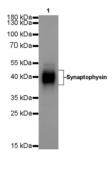
WB result of Synaptophysin Rabbit mAb Primary antibody: Synaptophysin Rabbit mAb at 1/500 dilution Lane 1: PC12 whole cell lysate 20 µg Secondary antibody: #JP20040 at 1/10000 dilution Predicted MW: 38 kDa Observed MW: 38~45 kDa Exposure time: 1.3s
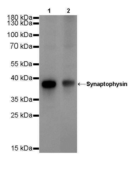
WB result of Synaptophysin Rabbit mAb Primary antibody: Synaptophysin Rabbit mAb at 1/500 dilution Lane 1: mouse cerebellum lysate 20 µg Lane 2: mouse brain lysate 20 µg Secondary antibody: #JP20040 at 1/10000 dilution Predicted MW: 38 kDa Observed MW: 38 kDa Exposure time: 1.3s
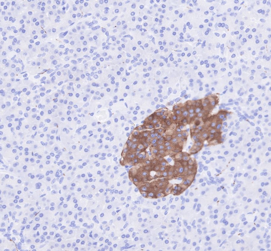
IHC shows positive staining in paraffin-embedded human pancreas. Anti-Synaptophysin antibody was used at 1/1000 dilution, Secondary antibody: #JP20040. Counterstained with hematoxylin. Heat mediated antigen retrieval with Tris/EDTA buffer pH9.0 was performed before commencing with IHC staining protocol.
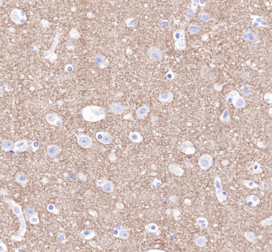
IHC shows positive staining in paraffin-embedded human brain. Anti-Synaptophysin antibody was used at 1/1000 dilution, Secondary antibody: #JP20040. Counterstained with hematoxylin. Heat mediated antigen retrieval with Tris/EDTA buffer pH9.0 was performed before commencing with IHC staining protocol.
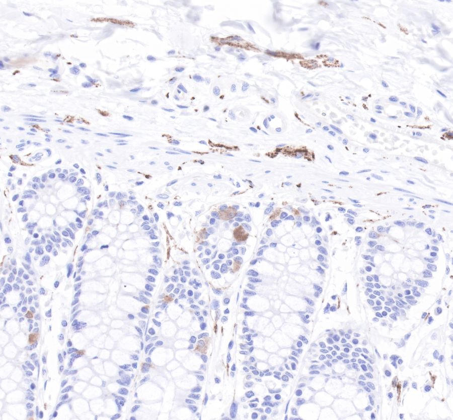
IHC shows positive staining in paraffin-embedded human colon. Anti-Synaptophysin antibody was used at 1/1000 dilution, Secondary antibody: #JP20040. Counterstained with hematoxylin. Heat mediated antigen retrieval with Tris/EDTA buffer pH9.0 was performed before commencing with IHC staining protocol.
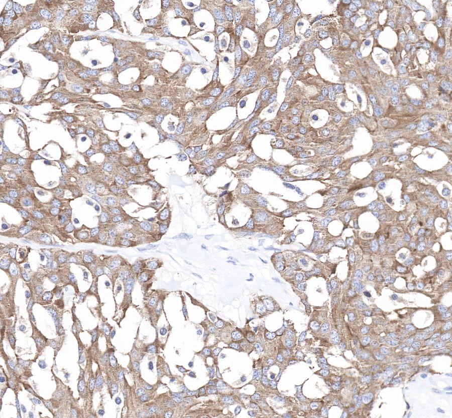
IHC shows positive staining in paraffin-embedded human medullary thyroid carcinoma. Anti-Synaptophysin antibody was used at 1/1000 dilution, Secondary antibody: #JP20040. Counterstained with hematoxylin. Heat mediated antigen retrieval with Tris/EDTA buffer pH9.0 was performed before commencing with IHC staining protocol.
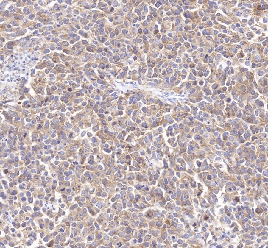
IHC shows positive staining in paraffin-embedded human small cell lung carcinoma. Anti-Synaptophysin antibody was used at 1/1000 dilution, Secondary antibody: #JP20040. Counterstained with hematoxylin. Heat mediated antigen retrieval with Tris/EDTA buffer pH9.0 was performed before commencing with IHC staining protocol.
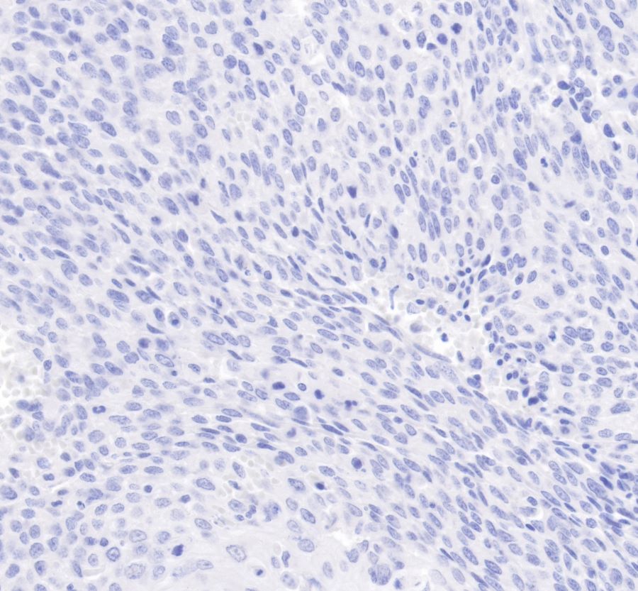
Negative tissue: IHC shows negative staining in paraffin-embedded human cervix cancer. Anti-Synaptophysin antibody was used at 1/1000 dilution, Secondary antibody: #JP20040. Counterstained with hematoxylin. Heat mediated antigen retrieval with Tris/EDTA buffer pH9.0 was performed before commencing with IHC staining protocol.
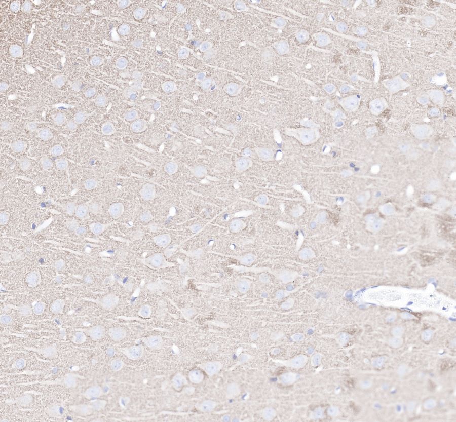
IHC shows positive staining in paraffin-embedded mouse brain. Anti-Synaptophysin antibody was used at 1/4000 dilution, Secondary antibody: #JP20040. Counterstained with hematoxylin. Heat mediated antigen retrieval with Tris/EDTA buffer pH9.0 was performed before commencing with IHC staining protocol.
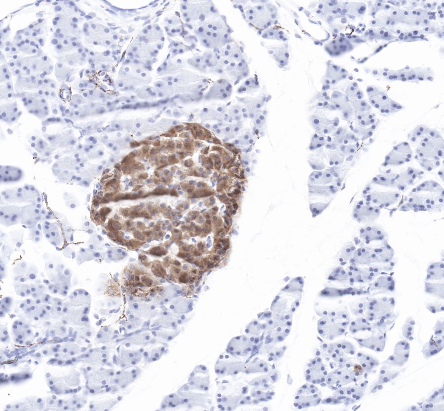
IHC shows positive staining in paraffin-embedded mouse pancreas. Anti-Synaptophysin antibody was used at 1/4000 dilution, Secondary antibody: #JP20040. Counterstained with hematoxylin. Heat mediated antigen retrieval with Tris/EDTA buffer pH9.0 was performed before commencing with IHC staining protocol.
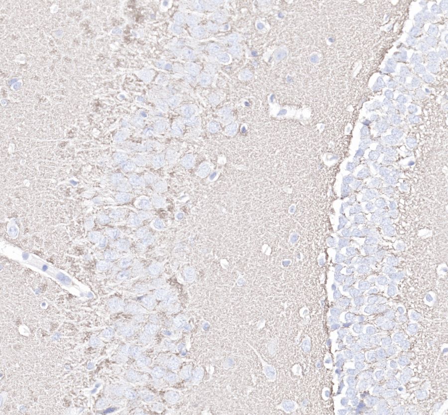
IHC shows positive staining in paraffin-embedded rat brain. Anti-Synaptophysin antibody was used at 1/4000 dilution, Secondary antibody: #JP20040. Counterstained with hematoxylin. Heat mediated antigen retrieval with Tris/EDTA buffer pH9.0 was performed before commencing with IHC staining protocol.
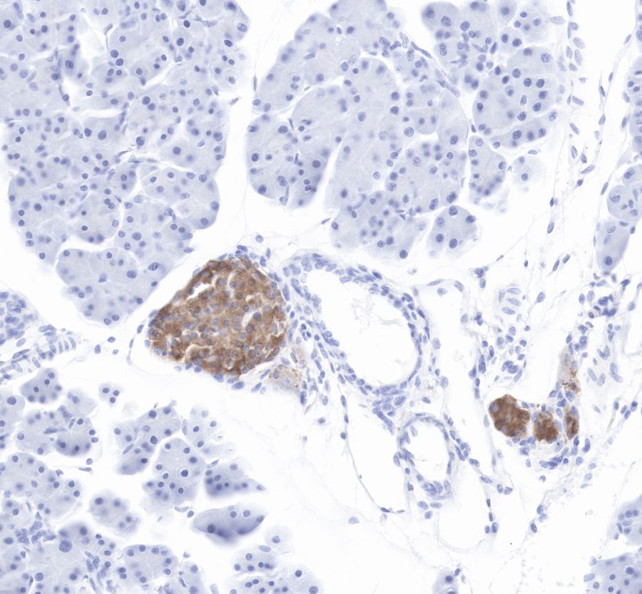
IHC shows positive staining in paraffin-embedded rat pancreas. Anti-Synaptophysin antibody was used at 1/4000 dilution, Secondary antibody: #JP20040. Counterstained with hematoxylin. Heat mediated antigen retrieval with Tris/EDTA buffer pH9.0 was performed before commencing with IHC staining protocol.
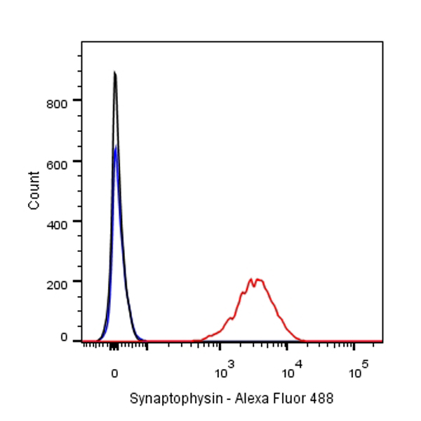
Flow cytometric analysis of PC-12 cells labelling Synaptophysin antibody at 1/500 (0.1ug) dilution/ (red) compared with a Rabbit monoclonal IgG (Black) isotype control and an unlabelled control (cells without incubation with primary antibody and secondary antibody) (Blue). Secondary antibody:#JP20025.