
Mouse anti-Erk1/2 Monoclonal Antibody(784CT7.6.3)描述别名宿主特异性反应种属应用分子量类型克隆号同种型储存/保存方法存储溶液研究领域背景说明细胞定位UniProt参考文献
| 概述 | |
| 描述 |
Mouse Monoclonal Antibody (Mab) |
| 别名 |
Erk1抗体;Mitogen-activated protein kinase 3; MAP kinase 3; MAPK 3; ERT2; Extracellular signal-regulated kinase 1; ERK-1; Insulin-stimulated MAP2 kinase; MAP kinase isoform p44; p44-MAPK; Microtubule-associated protein 2 kinase; p44-ERK1; MAPK3; ERK1; PRKM3
|
| 宿主 |
Mouse
|
| 特异性 |
Purified His-tagged Erk1/2 protein was used to produced this monoclonal antibody.
|
| 反应种属 |
Human, Mouse
|
| 应用 |
FC~~1:25
WB~~1:1000 |
| 分子量 |
Predicted molecular weight: 43kD
Disclaimer note: The observed molecular weight of the protein may vary from the listed predicted molecular weight due to post translational modifications, post translation cleavages, relative charges, and other experimental factors. |
| 性能 | |
| 类型 |
Monoclonal Antibody
|
| 克隆号 |
784CT7.6.3
|
| 同种型 |
IgG2a
|
| 储存/保存方法 |
Maintain refrigerated at 2-8°C for up to 2 weeks. For long term storage store at -20°C in small aliquots to prevent freeze-thaw cycles.
|
| 存储溶液 |
Purified monoclonal antibody supplied in PBS with 0.09% (W/V) sodium azide. This antibody is purified through a protein G column, eluted with high and low pH buffers and neutralized immediately, followed by dialysis against PBS.
|
| 研究领域 |
Cancer;Cell Biology;Immunology;Neuroscience;Signal Transduction
|
| 靶标 | |
| 背景说明 |
Serine/threonine kinase which acts as an essential component of the MAP kinase signal transduction pathway. MAPK1/ERK2 and MAPK3/ERK1 are the 2 MAPKs which play an important role in the MAPK/ERK cascade. They participate also in a signaling cascade initiated by activated KIT and KITLG/SCF. Depending on the cellular context, the MAPK/ERK cascade mediates diverse biological functions such as cell growth, adhesion, survival and differentiation through the regulation of transcription, translation, cytoskeletal rearrangements. The MAPK/ERK cascade plays also a role in initiation and regulation of meiosis, mitosis, and postmitotic functions in differentiated cells by phosphorylating a number of transcription factors. About 160 substrates have already been discovered for ERKs. Many of these substrates are localized in the nucleus, and seem to participate in the regulation of transcription upon stimulation. However, other substrates are found in the cytosol as well as in other cellular organelles, and those are responsible for processes such as translation, mitosis and apoptosis. Moreover, the MAPK/ERK cascade is also involved in the regulation of the endosomal dynamics, including lysosome processing and endosome cycling through the perinuclear recycling compartment (PNRC); as well as in the fragmentation of the Golgi apparatus during mitosis. The substrates include transcription factors (such as ATF2, BCL6, ELK1, ERF, FOS, HSF4 or SPZ1), cytoskeletal elements (such as CANX, CTTN, GJA1, MAP2, MAPT, PXN, SORBS3 or STMN1), regulators of apoptosis (such as BAD, BTG2, CASP9, DAPK1, IER3, MCL1 or PPARG), regulators of translation (such as EIF4EBP1) and a variety of other signaling-related molecules (like ARHGEF2, FRS2 or GRB10). Protein kinases (such as RAF1, RPS6KA1/RSK1, RPS6KA3/RSK2, RPS6KA2/RSK3, RPS6KA6/RSK4, SYK, MKNK1/MNK1, MKNK2/MNK2, RPS6KA5/MSK1, RPS6KA4/MSK2, MAPKAPK3 or MAPKAPK5) and phosphatases (such as DUSP1, DUSP4, DUSP6 or DUSP16) are other substrates which enable the propagation the MAPK/ERK signal to additional cytosolic and nuclear targets, thereby extending the specificity of the cascade.
|
| 细胞定位 |
Cytoplasm. Nucleus. Note=Autophosphorylation at Thr-207 promotes nuclear localization
|
| UniProt |
P27361
|
| 参考文献 | |
| 参考文献 |
Sano H., et al. J. Biol. Chem. 277:19439-19447(2002).
Ronnstrand L., et al. Cell. Mol. Life Sci. 61:2535-2548(2004). Charest D.L., et al. Mol. Cell. Biol. 13:4679-4690(1993). Aebersold D.M., et al. Submitted (APR-2001) to the EMBL/GenBank/DDBJ databases. Cheng H., et al. Submitted (FEB-2006) to the EMBL/GenBank/DDBJ databases. |
实验结果图
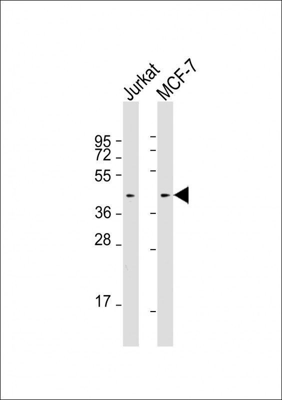
All lanes : Anti-Erk1/2 Antibody at 1:4000 dilutionLane 1: Jurkat whole cell lysatesLane 2: MCF-7 whole cell lysatesLysates/proteins at 20 μg per lane. SecondaryGoat Anti-mouse IgG, (H+L), Peroxidase conjugated at 1/10000 dilution. Predicted band size : 41 kDaBlocking/Dilution buffer: 5% NFDM/TBST.
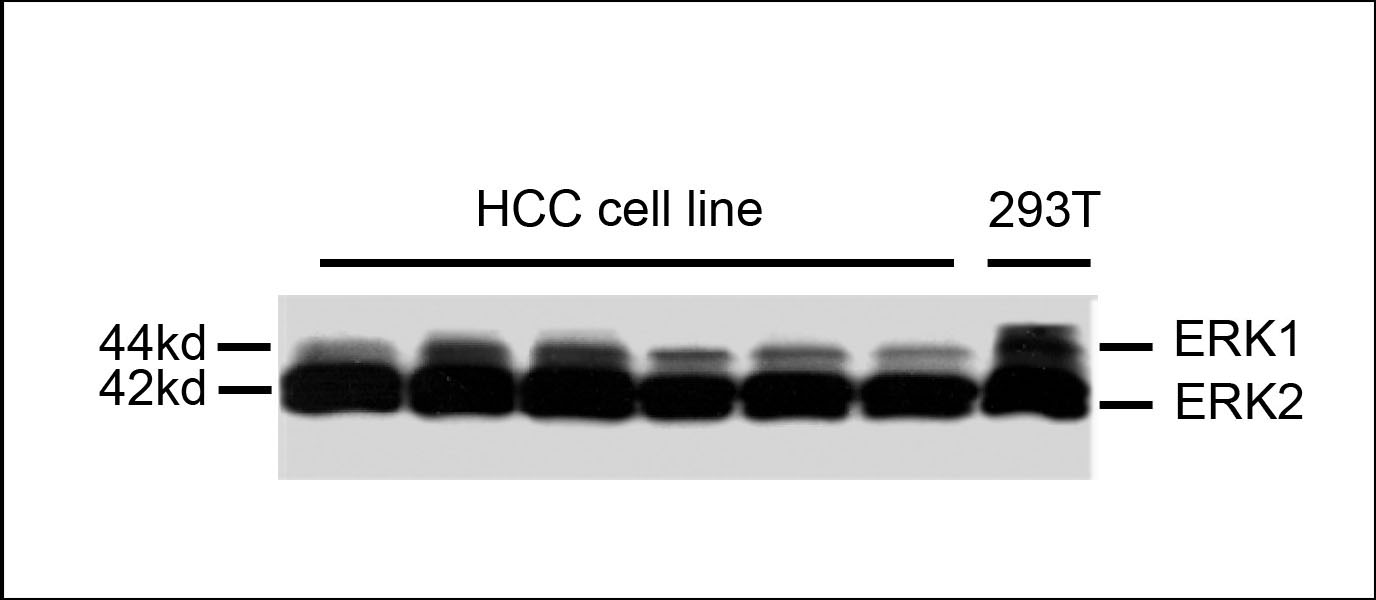
Western blot analysis of extracts from HCC cell line and 293T cells, using mouse monoclonal antibody Erk1/2 Antibody (Cat. #JP100329).
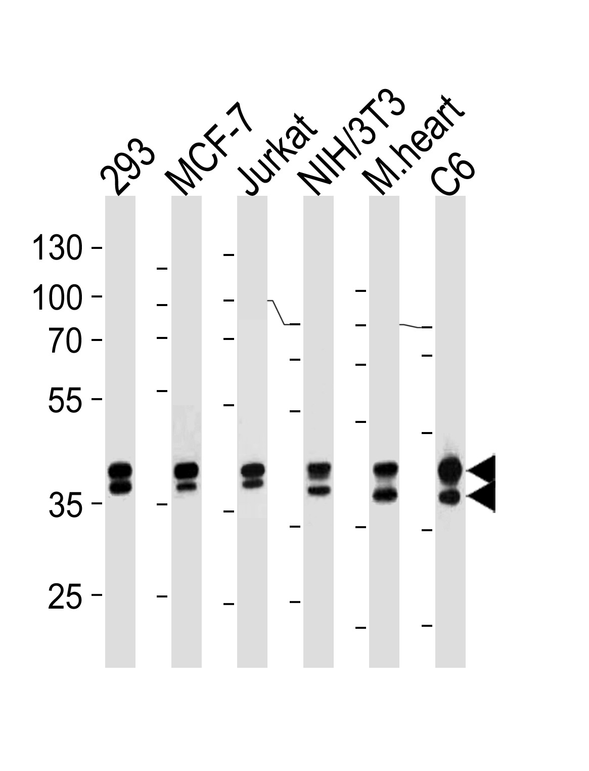
Erk1/2 Antibody (Cat. #JP100329) western blot analysis in 293,MCF-7,Jurkat,mouse NIH/3T3,rat C6 cell line and mouse heart lysates (35μg/lane).This demonstrates the Erk1/2 antibody detected the Erk1/2 protein (arrow).